
Industrial
Industrial
Sumérjase en artículos detallados y seminarios web centrados en la inspección eficaz, los flujos de trabajo optimizados y la comodidad ergonómica en contextos industriales y patológicos. Los temas tratados incluyen el control de calidad, el análisis de materiales y la microscopía en patología, entre muchos otros. Este es el lugar donde obtendrá información valiosa sobre el uso de tecnologías de vanguardia para mejorar la precisión y la eficacia de los procesos de fabricación, así como el diagnóstico y la investigación patológicos precisos.

Differential Interference Contrast (DIC) Microscopy
This article demonstrates how differential interference contrast (DIC) can be actually better than brightfield illumination when using microscopy to image unstained biological specimens.
 taken with a ring light (RL) and near vertical illumination (NVI).")
Microscope Illumination for Industrial Applications
Inspection microscope users can obtain information from this article which helps them choose the optimal microscope illumination or lighting system for inspection of parts or components.
 cells taken with phase contrast.")
Phase Contrast and Microscopy
This article explains phase contrast, an optical microscopy technique, which reveals fine details of unstained, transparent specimens that are difficult to see with common brightfield illumination.

How to Prepare your Specimen for Immunofluorescence Microscopy
Immunofluorescence (IF) specimen preparations can be analyzed by various fluorescence microscopy techniques, depending on the research application. IF is indispensable for researchers using a simple…

Microscopios de campo oscuro
El método de contraste de campo oscuro aprovecha la difracción o dispersión de la luz de las estructuras de un espécimen biológico o las características no uniformes de una muestra de material.

A Guide to Phase Contrast
A phase contrast light microscope offers a way to view the structures of many types of biological specimens in greater contrast without the need of stains.

Introduction to Widefield Microscopy
This article gives an introduction to widefield microscopy, one of the most basic and commonly used microscopy techniques. It also shows the basic differences between widefield and confocal…

Photoactivatable, Photoconvertible, and Photoswitchable Fluorescent Proteins
Fluorescent proteins (FPs) such as GFP, YFP or DsRed are powerful tools to visualize cellular components in living cells. Nevertheless, there are circumstances when classical FPs reach their limits.…
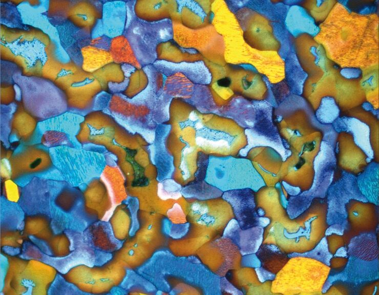
Metallography with Color and Contrast
The examination of microstructure morphology plays a decisive role in materials science and failure analysis. There are many possibilities of visualizing the real structures of materials in the light…
