
Especialidades médicas
Especialidades médicas
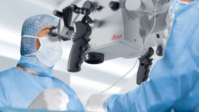
Explore una completa colección de recursos científicos y clínicos adaptados a los profesionales sanitarios, que incluye opiniones de colegas, estudios de casos clínicos y simposios. Diseñada para neurocirujanos, oftalmólogos y especialistas en cirugía plástica y reparadora, otorrinolaringología y odontología. Esta colección destaca los últimos avances en microscopía quirúrgica. Descubra cómo las tecnologías quirúrgicas de vanguardia, como la fluorescencia AR, la visualización 3D y las imágenes OCT intraoperatorias, permiten tomar decisiones con confianza y precisión en cirugías complejas.

A New Method for Convenient and Efficient Multicolor Imaging
The technique combining hyperspectral unmixing and phasor analysis was developed to simplify the process of getting images from a sample labeled with multiple fluorophores. This aggregate method…

Considerations for Multiplex Live Cell Imaging
Simultaneous multicolor imaging for successful experiments: Live-cell imaging experiments are key to understand dynamic processes. They allow us to visually record cells in their living state, without…

How to Select the Right Solution for Visual Inspection
This article helps users with the decision-making process when selecting a microscope as a solution for routine visual inspection. Important factors that should be considered are described.

How to Use a Digital Microscope to Streamline Inspection Processes
Watch this webinar for inspiration and expert advice on how to make quality control simpler, quicker, and easier. Learn how to perform comprehensive visual inspection, including comparison,…
Neuroscience Images
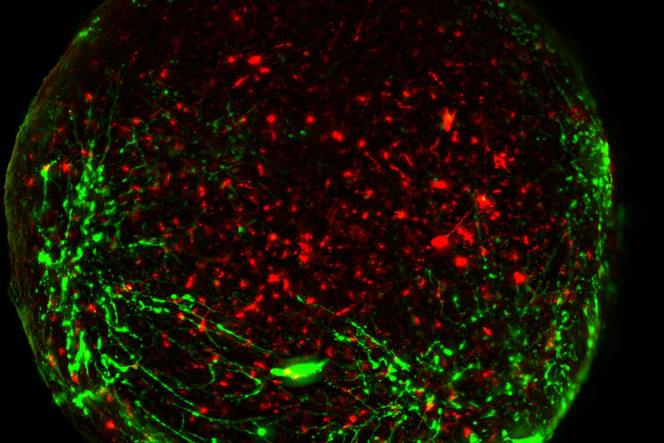
Neuroscience commonly uses microscopy to study the nervous system’s function and understand neurodegenerative diseases.

Workflows and Instrumentation for Cryo-electron Microscopy
Cryo-electron microscopy is an increasingly popular modality to study the structures of macromolecular complexes and has enabled numerous new insights in cell biology. In recent years, cryo-electron…
Organismos Modelo en la Investigación
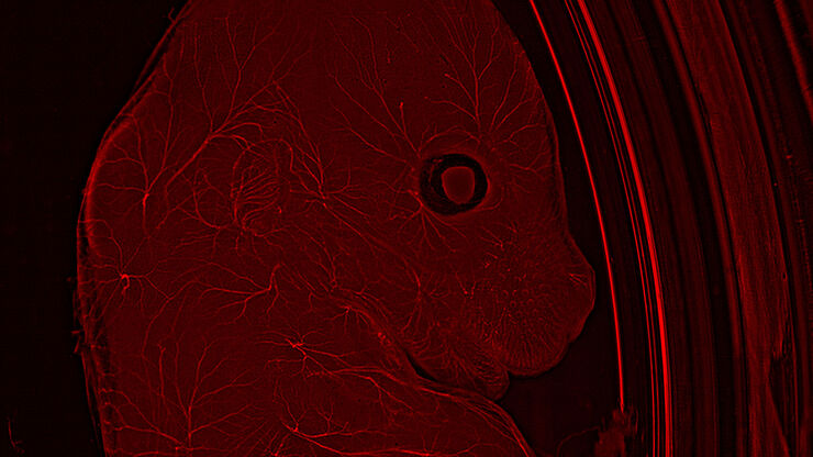
Un organismo modelo es una especia empleada por los investigadores para estudiar procesos biológicos específicos. Sus características genéticas son similares a las de los humanos y se emplean de forma…

Microscopy in Virology
The coronavirus SARS-CoV-2, causing the Covid-19 disease effects our world in all aspects. Research to find immunization and treatment methods, in other words to fight this virus, gained highest…
Investigación del cáncer
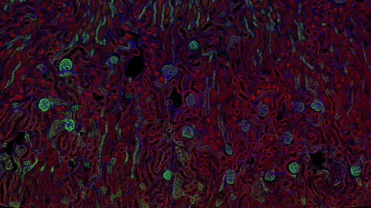
El cáncer es una enfermedad compleja y heterogénea causada por células con una regulación deficiente del crecimiento. Los cambios genéticos y epigenéticos en una célula o en un grupo de ellas alteran…


