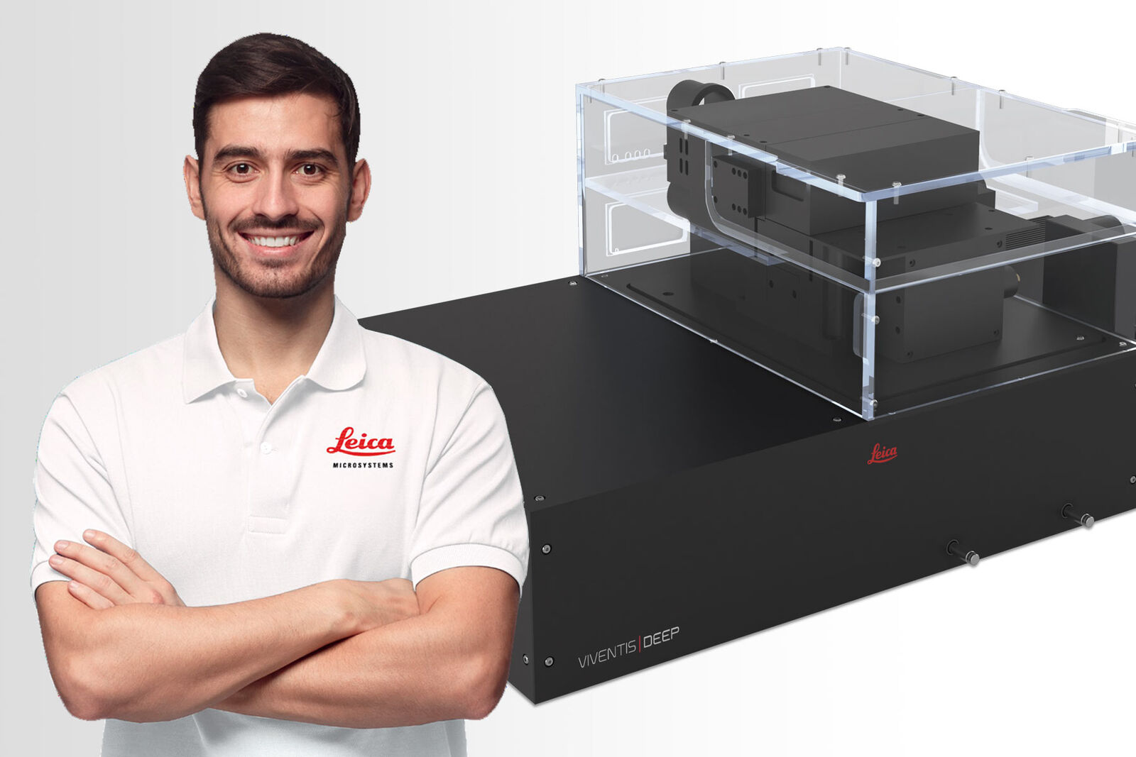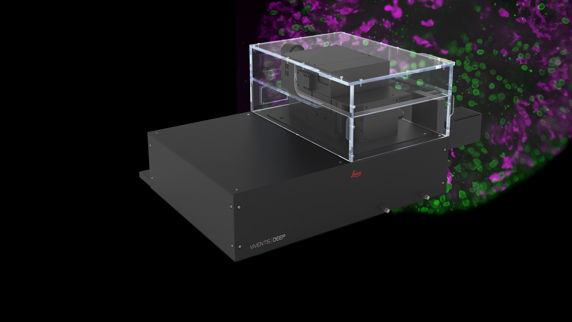
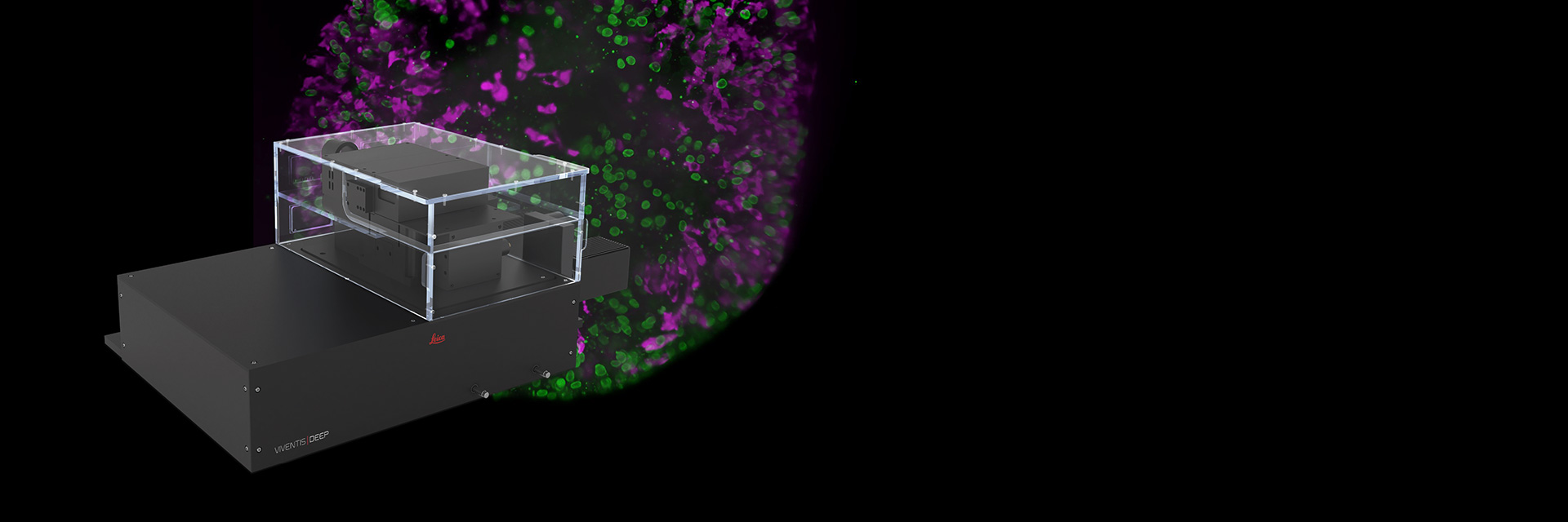
Viventis Deep Dual View Light Sheet Microscope
Image live and cleared samples with the Viventis Deep light sheet system to unveil intricate details and dynamic processes of biological systems. Its unique combination of multi-well and multi-position capabilities helps you to capture life in its entirety as it develops. By simply switching the replaceable optics, you can adapt the system for cleared samples to gain deeper context.
Popular Configurations

Loading...
|

Loading...
|

Loading...
|

Loading...
|

Loading...
|

Loading...
|

Loading...
|

Loading...
|

Loading...
|
Don’t see your Configuration? Request for individual Quote
Show popular configurations
Explore and buy pre-configured microscopy solutions in our online shop. Enjoy a seamless online shopping experience.
Explore life in depth
The Viventis Deep microscope helps you to expand the spatio-temporal understanding of your sample to its full depth, thanks to increased spatio-temporal resolution.
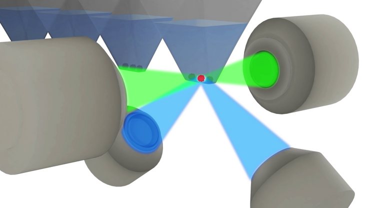
Achieve detailed volumetric imaging for a complete view of the sample with a patented combination of
- Dual illumination
- Dual view detection
- Multi-position
- Open top sample holder
You can even image large light scattering samples over time with outstanding quality for meaningful downstream analysis, while minimizing light dose and maintaining sample accessibility.

Explore life events with long-term imaging
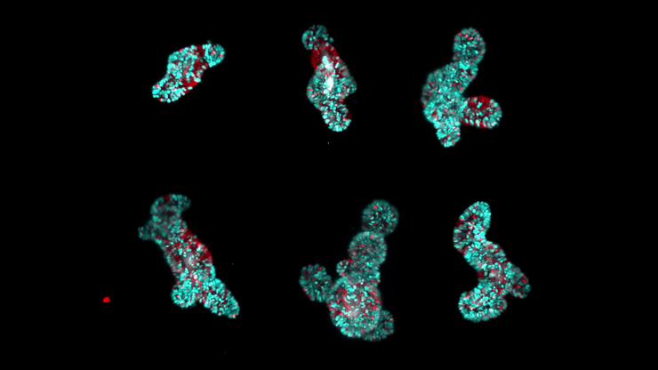
In a variety of model systems, including intestine, liver and human colon cancer organoids and zebrafish embryos, the gentle light sheet technology of the Viventis Deep microscope provides high image quality while preserving sample viability.
In addition, the advanced incubation solution and easy media exchange even during a running experiment preserves physiological conditions. Easily handle even more complex experimental conditions. To study responses and to ensure specific results, you can add drugs and use an optional photomanipulation arm during a running time-lapse.
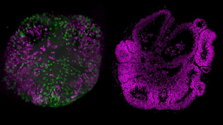
Explore live and cleared samples flexibly
The combination of live and fixed or cleared imaging experiments supports capturing both dynamic information and molecular context. Viventis Deep enables imaging of live and cleared samples in a single system by exchanging the detection optics.
The cleared-sample module accommodates diverse clearing media and refractive indices and supports samples from hundreds of micrometers up to 9x6x6 mm. Tiling, stitching, and computational clearing enhance workflows for imaging large, cleared samples, and help maintain crisp image quality throughout.
Shape the light sheet technology to fit your needs
The Viventis Deep microscope helps you to tune the thickness of the scanned Gaussian beam (DLSM). You can tailor the light sheet with a focus on resolution or field of view.
Its flexible software allows you to change settings while the timelapse is running. In addition, a Python application programming interface (API) lets you code and plug in custom macros for your own experimental ideas.
To prevent your sample from moving out of the field of view (FOV) during time-lapse, intelligent online object tracking changes the stage coordinates.
Enabling your success with complete workflow support
Keep your operations running around the globe with best-in-class services entirely dedicated to microscopy and over 175 years of history.
Key features
- Leica Team: 500+ Service & Application experts
- Leica Training: 4-level factory certification program
- Leica Logistics: 5 regional hubs for genuine parts
- Leica OneCall: PhD-level hotline assistance