Integrated Serial Sectioning and Cryo-EM Workflows for 3D Biological Imaging
A practical walkthrough of integrated electron microscopy workflows using UC Enuity and Aivia

This on-demand webinar explores how integrated tools can support electron microscopy workflows from sample preparation to image analysis. Experts Andreia Pinto, Adrian Boey, and Hoyin Lai present the UC Enuity ultramicrotome and Aivia image analysis platform, and demonstrate how these tools can be applied in both room temperature and cryogenic environments. The session includes practical examples in array tomography, deep learning-based segmentation, and cryo-lift-out workflows for biological imaging.
Key learnings
- How to streamline serial sectioning for volume EM using the UC Enuity and AT module
- How to achieve in-situ targeting under cryogenic conditions for cryo-EM workflows
- How integrated imaging and AI-powered analysis enhance 3D EM data interpretation
Integrated automation, AI-driven image analysis, and cryogenic techniques for precise 3D reconstruction
Gain a clear understanding of how to streamline your electron microscopy (EM) workflow using Leica Microsystems’ UC Enuity and Aivia platforms. Start by exploring how UC Enuity, a motorized ultramicrotome, supports consistent and efficient ultramicrotomy. Features like auto-alignment and auto-trimming help reduce manual effort and improve reproducibility—especially valuable when working with array tomography, where maintaining section integrity is essential for accurate 3D reconstruction.
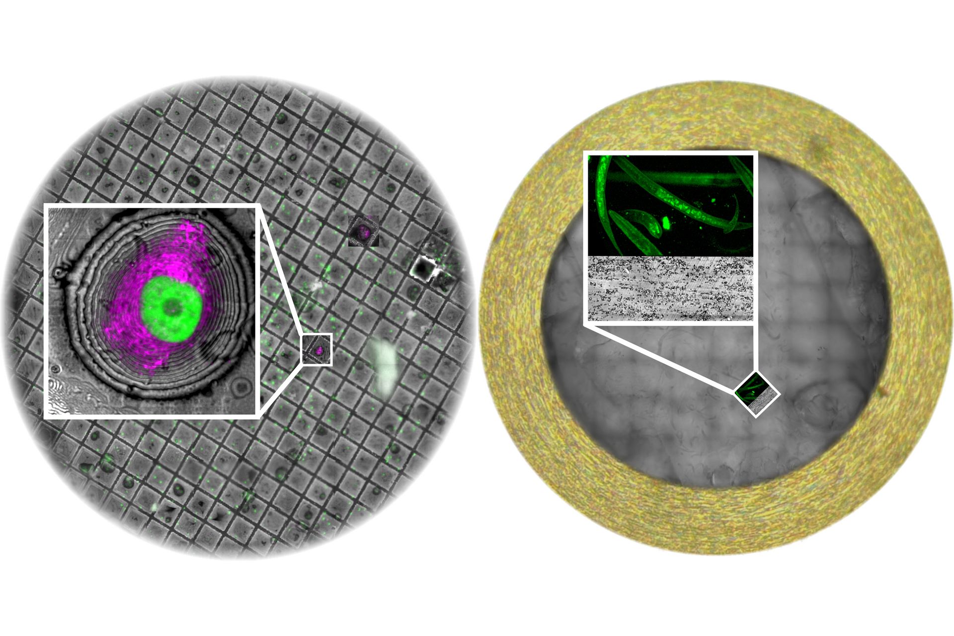
See how these tools are applied in a real-world context through the preparation of Trichomonas parasite samples. Learn how to use the OTO staining method to enhance contrast and prepare ribbons of ultrathin sections for downstream imaging.
Next, dive into image analysis with Aivia, Leica’s AI-powered platform. Discover how to train deep learning models using annotated EM data to segment cells and organelles. Use 3D object reconstruction and mesh editing tools to visualize structures such as flagella, hydrogenosomes, nuclei, and lysosomes. Explore how annotated data can be reused for future model training and how to extract quantitative insights into organelle morphology and localization.
Expand your workflow into cryogenic applications by integrating UC Enuity into cryo sample preparation. Learn how cryo-planing techniques expose vitrified biological material for imaging, and how to use the UC Enuity cryo configuration and STELLARIS Cryo to identify regions of interest through fluorescence microscopy. Follow the full cryo lift-out process—from sample freezing and fluorescence targeting to Cryo-FIB SEM and Cryo-TEM imaging.
Case studies with C. elegans and mouse brain tissue illustrate how to locate and image specific structures in complex biological samples. See how to correlate confocal and SEM data, prepare lamellae, and perform 3D reconstructions—even when working with challenging conditions like surface artifacts or ice contamination.
This session offers practical insights into combining automation, AI, and cryo techniques to support structural and cellular biology research.
Related Articles
-
, insulin SGs (orange), microtubules (red), nucleus (yellow), and plasma membrane (transparent).")
High-Pressure Freezing Protocols for Ultrastructural 3D EM
High pressure freezing (HPF) can help preserve hydrated cells and tissues close to their biological…
Mar 31, 2026Read article -
Ultramicrotome UC Enuity in Practice: Stable 15 nm Sections at ZFE
After using the UCT and UC6 ultramicrotomes, Claudia Mayrhofer calls UC Enuity a leap in…
Mar 25, 2026Read article -

Ultramicrotomy eBook: Targeting, Trimming & Alignment
Ultramicrotomy is evolving rapidly, and today’s microscopes demand high‑quality sections, precise…
Feb 11, 2026Read article
