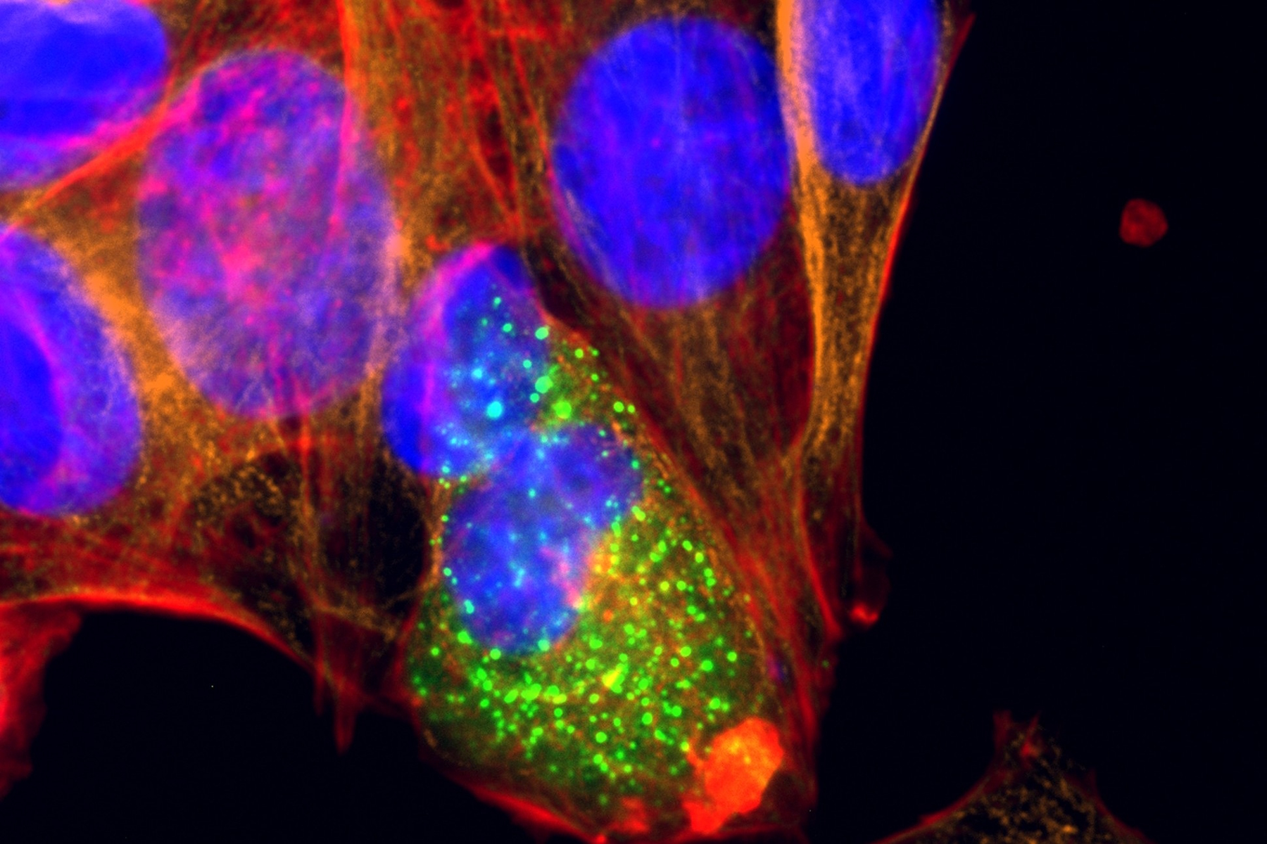
 and co-stained for nuclear DNA (Hoechst 33342), microtubules (Alexa 555) and F-actin (ATTO 643). Image was captured on Mateo FL.")
"This resource is a result of a collaboration between Leica Microsystems and Bitesize Bio, created to help you understand the intersection of 2D cell culture and modern imaging technologies. We hope you find it both accessible and applicable to your research.
Our aim is to share practical knowledge that helps scientists overcome real-life challenges at the bench.
2D cell culture may be familiar, but getting the most out of your cultures increasingly depends on understanding microscopy and imaging—areas that can feel like someone else’s specialty.
However, you may find that by learning just a little more about microscopy technologies, you can expand what’s possible in your experiments. The right imaging approach can help you answer new biological questions, generate more reliable data, and improve the efficiency of routine steps you already perform, such as checking confluency, monitoring transfections, or counting cells.
We wrote this eBook to provide you with the tools to bring microscopy and AI analysis to your 2D cell culture with confidence. In the chapters ahead, we explore both traditional and digital imaging techniques,alongside AI-powered tools that simplify complex analysis tasks, and improve data documentation. By combining these technologies with your existing cell culture expertise, you can bring greater consistency,reproducibility, and confidence to your work."
Skye Marshall and Thomas Warwick
Editors, Bitesize Bio
