Tissue imaging with STELLARIS confocal platform
The STELLARIS confocal platform gives you access to multiphoton imaging, STED super-resolution, fluorescence lifetime imaging microscopy (FLIM), and Digital Light Sheet (DLS), to image the most demanding tissue samples. This gallery shows images from tissue obtained with the STELLARIS confocal platform.
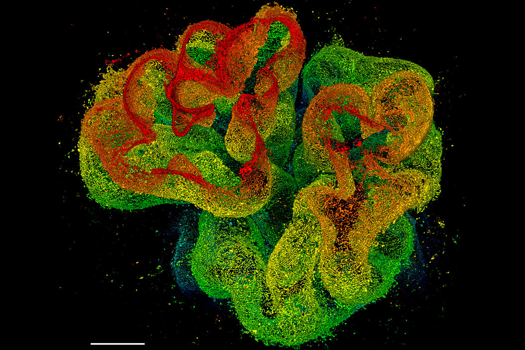
3D STED 775 deep nanoscopy of glomerulus in cleared kidney tissue

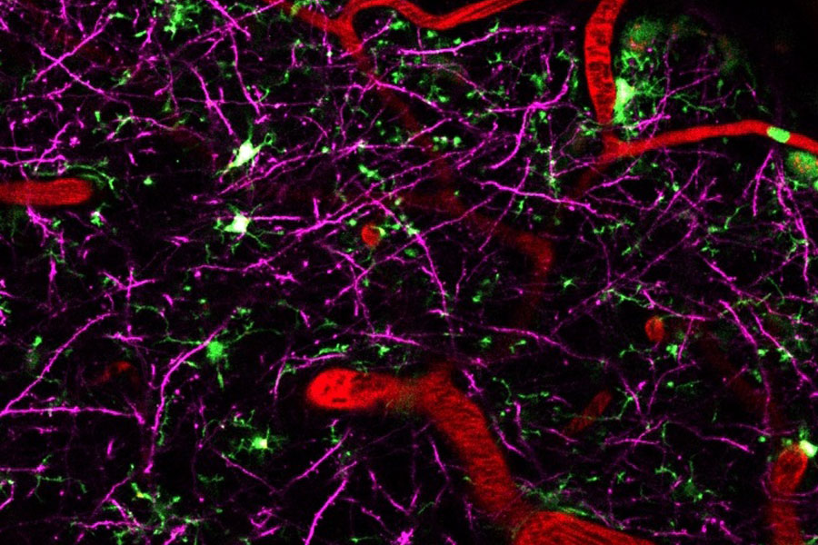
4 colour live imaging of mouse brain cortex
Neurodegenerative diseases are usually caused by a combination of different factors. Being able to visualize multiple players at once greatly helps to investigate and understand how different compoonents interact and influence each other. To do this, a microscope that combines deep tissue penetration with spectral flexibility to enable multicolor imaging liek STELLARIS 8 DIVE is the best choice.

Multicolor Deep In Vivo Imaging to uncover the interplay of various players in neurodegenerative diseases
Neurodegenerative diseases are usually caused by a combination of different factors. Being able to visualize multiple players at once greatly helps to investigate and understand how different compoonents interact and influence each other. To do this, microscope that combines deep tissue penetration with spectral flexibility to enable multicolor imaging like STELLARIS 8 DIVE is the best choice.
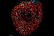

, actin network (ATTO 647N), and nuclear pore basket (CF 680R).")