Neurosurgery Microscopy Solutions
Leica Microsystems offers a range of surgical microscope solutions (PROvido, M530 OHX, ARveo 8) to support neurosurgeons during the most complex microsurgery applications. Our cutting-edge neurosurgery microscopes provide exclusive optical technology, including built-in fluorescence filters, Augmented Reality (AR) capabilities, and 3D visualization.
They are designed to address surgeons’ needs, offering brilliant illumination, upgradability, and adjustable viewing accessories to support ergonomics. Our solutions for neurosurgery enable surgeons to see more detailed information, facilitating more confident, real-time surgical decisions and leading to clinical value creation.
Contact a local specialist for expert advice on the right solution for your needs and budget.
Neurosurgery microscopy solutions

Rely on proven Leica surgical visualization and illumination features enhanced with new digital capabilities, such as the 3D Augmented Reality fluorescence applications: GLOW800 for vascular neurosurgery.

Experience surgical workflow efficiency by easily connecting to compatible surgical devices. Further augment your microscope view intraoperatively with data from other sources, such as leading IGS systems, endoscopic video feeds and optimized data processing as well as connectivity for DICOM.

Share surgeries live in 4K and 3D with your entire team to optimize teaching and collaboration. Choose from a variety of visualization options, such as the MyVeo surgical headset, available for the evolved ARveo 8 digital visualization microscope for neurosurgery.
Applications of Neurosurgery Operating Microscopes
Neurosurgeons rely on surgical microscopes to visualize the operating field and fine anatomical details of brain structures in order to perform a wide range of surgical procedures with high precision.
Applications of neurosurgery operating microscopes can include the following:
- Brain aneurysm clipping
- Tumor resections
- Arteriovenous malformation (AVM) treatment
- Cerebral artery bypass surgery
- Spine surgery
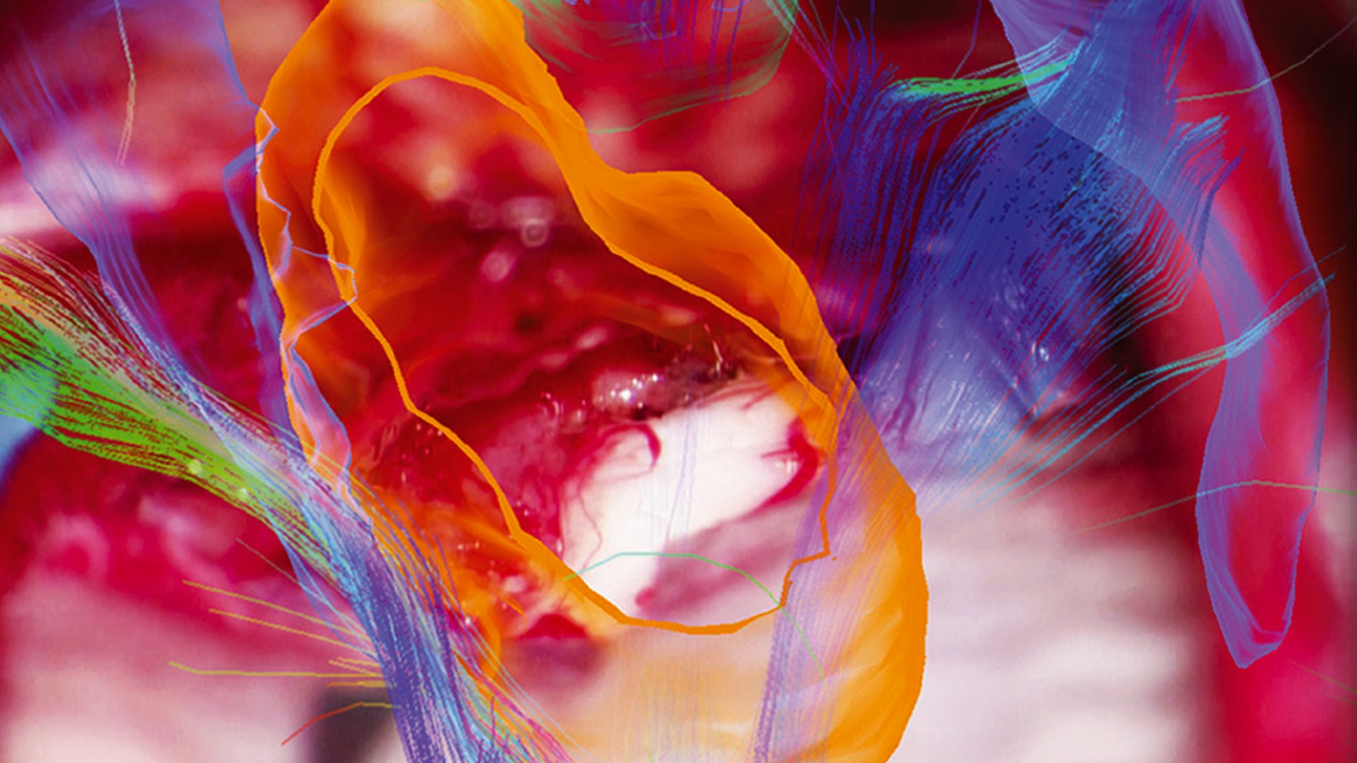
Leica microscopes for neurosurgery feature innovative, integrable technologies that enhance the surgeon's vision through Augmented Reality (AR). In vascular surgery, for example, GLOW800 AR fluorescence allows the neurosurgeon to clearly visualize blood flow in real-time and in 3D.
Challenges of Neurosurgery Microscopy
Neurosurgeons operate in deep and narrow cavities, while navigating through delicate tissue, locating anatomical structures with vitally important functions.
Achieving sufficient illumination and depth of field, as well as a large enough and unobstructed field of view, are some of the main challenges of using microscopy in neurosurgery.
Simultaneous and clear visualization of blood flow in neurovascular procedures, as well as differentiating between different tissue types in tumor resection surgery, are equally challenging in modern neurosurgery.
Finally, poor ergonomics resulting in musculoskeletal pain continues to be a major problem for neurosurgeons, impacting their quality of life and career longevity.
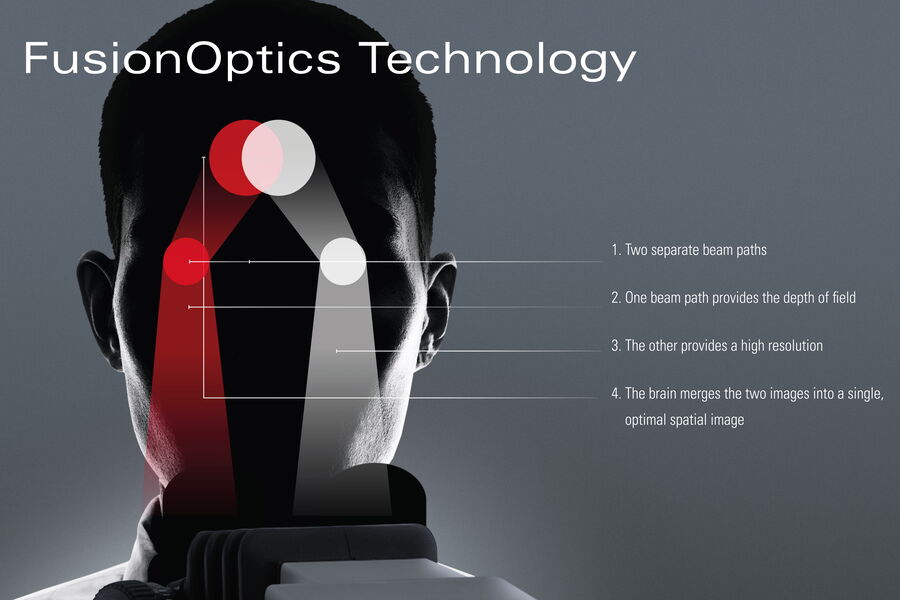
Advanced Optical Technology
The Leica microscopes for neurosurgery provide world-renowned optical quality with FusionOptics technology and state-of-the-art illumination for brilliant images and increased depth perception in deep cavities and include:
- Bright 400-Watt xenon lights available on the ARveo 8 and M530 OHX microscopes
- Bright 300-Watt xenon lights available on the PROvido microscope
- Small Angle Illumination (SAI) for optimal views to the bottom of deep, narrow cavities
- Apochromatic optics that deliver a clear, sharply focused image
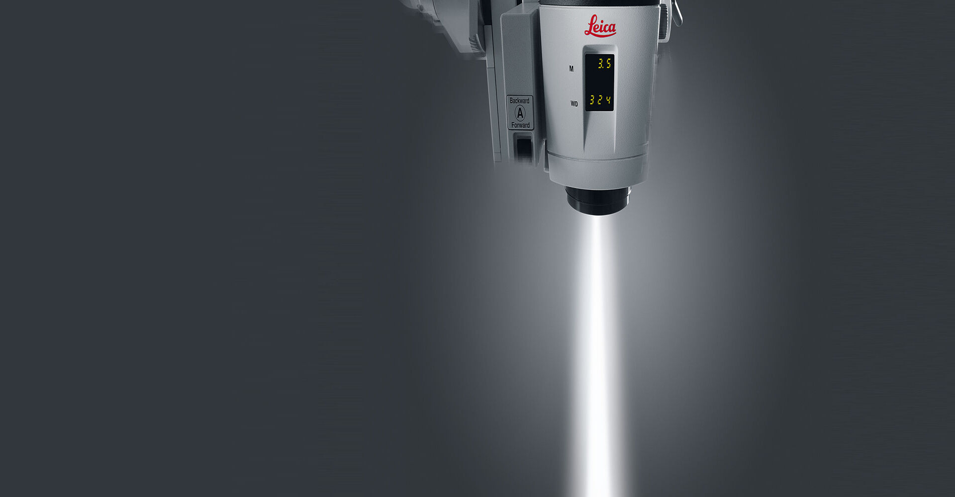
- SpeedSpot laser or Video AutoFocus to enable quick and easy focusing for the main surgeon, assistant(s) and video camera
- Leica Magnification Multiplier with 40% additional optical magnification (optional)
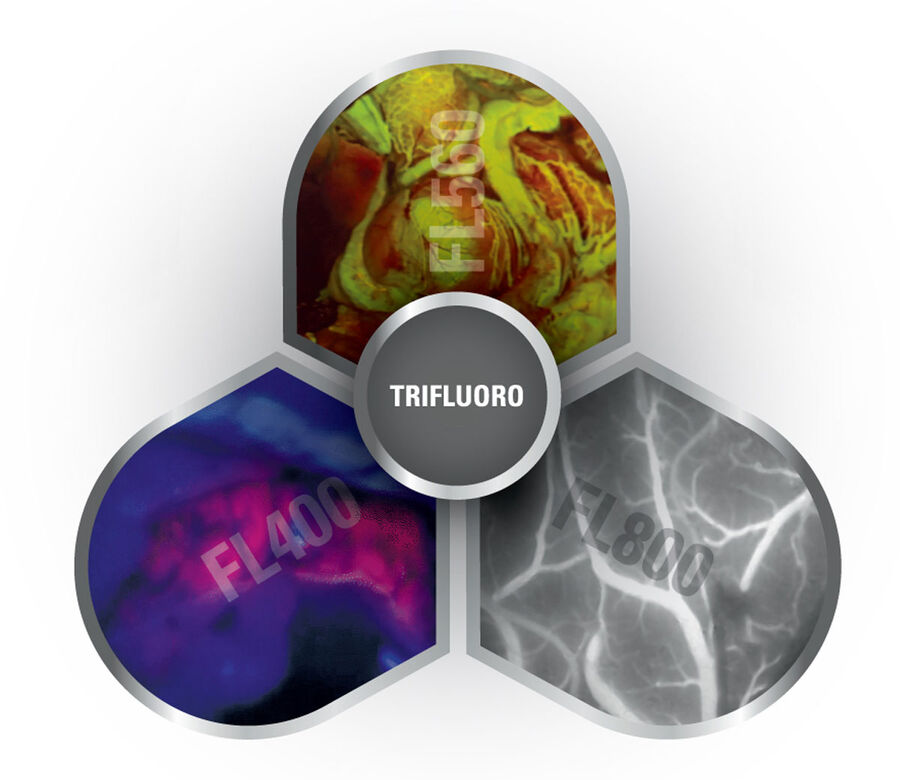
Enhanced View with Fluorescence Filters
Enhanced view with Fluorescence filters
Leica Microsystems is a pioneer in the field of fluorescence microscopy technology, developing innovative fluorescent modules for various medical applications such as:
- FL560 fluorescence: Get a single, real-time view of fluorescent and non-fluorescent areas with clear differentiation and high contrast during aneurysm clipping
- FL400 oncological fluorescence: When used with 5-aminolevulinic acid (5-ALA) this filter supports resection procedures by allowing differentiation of tumor tissue from healthy brain tissue
- FL800 intra-operative video-angiography module: When used in conjunction with ICG fluorescent dye, this feature allows neurosurgeons to clearly visualize blood flow in 2D.
Please check with your local Leica Microsystems representative for product registration status.
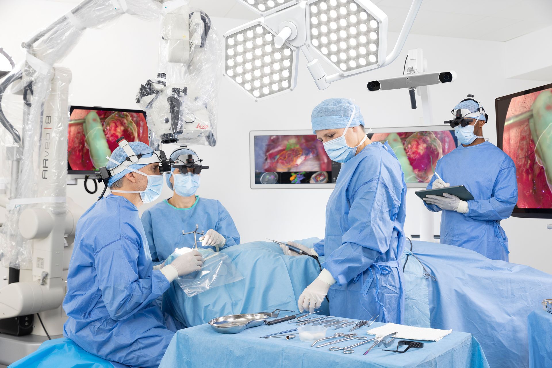
Augmenting Insights with Neurosurgical Microscopes
During delicate and complex neurosurgery procedures, one of the most significant challenges is to optimally preserve healthy brain tissue. Image-guided surgery (IGS) and robotic control in neurosurgical interventions facilitate intraoperative surgical decision-making and help surgeons achieve optimal patient outcomes.
Leica neurosurgical microscopes are compatible with leading neuro-navigation systems. These allow image injection of IGS systems directly into the surgeon's eyepieces or onto a 3D 4K screen, creating an augmented surgical view.
Learn about the Brainlab microscope navigation software, which is compatible with the Arveo neurosurgery microscope from Leica Microsystems, courtesy of BrainLab AG.
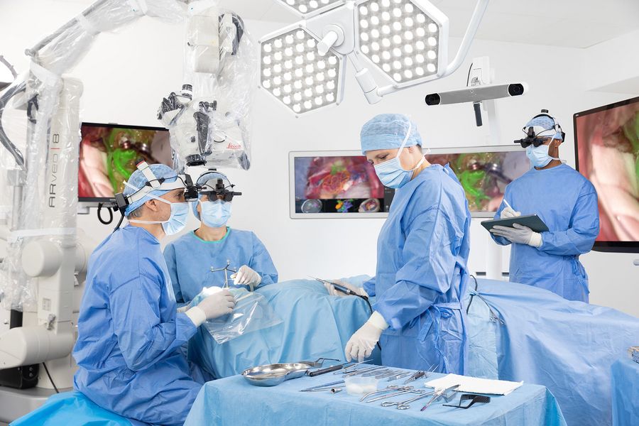
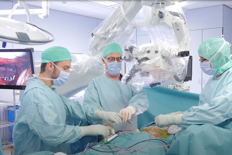
Visualization freedom of Leica Neurosurgery Operating Microscopes
There are a range of visualization options to ensure that surgeons have ease of movement and collaborate with their OR colleagues. The Evolved ARveo 8 surgical microscope combines a programmable footswitch and flexible handles. Leica neurosurgery microscopes provide great manoeuvrability, allowing for optimal positioning and enhanced ergonomics for both the surgeon and the resident during lengthy procedures.
The Evolved ARveo 8 surgical microscope combines optical and digital technologies with three interchangeable viewing options for the surgeon:
- Traditional oculars: These adaptable binoculars can be positioned and angled to meet the surgeon’s specific operating requirements
- Heads-up monitors: Heads-up displays enable everyone in the OR to see the same image as the surgeon on a 4K screen in 3D, improving training and documentation
- Surgical visualization headset: The MyVeo headset frees the surgeon from the microscope and external monitors by unifying essential clinical data in real-time directly in front of their eyes. Surgeons can perform exoscopic surgery using the surgical headset and 3D heads-up display.
Integration with Other Devices and Technologies
There are a range of accessories compatible with Leica neurosurgery microscope solutions. For example, the following advanced features of Evolved ARveo 8 neurosurgical microscope meet the needs of neurosurgeons for increased efficiency, uninterrupted workflow and augmented insights:
- Augmented Reality fluorescence applications by the GLOW AR Platform
- Fluorescence filters FL560, GLOW800
- IGS system integration of leading neuro-navigation providers
- Robotic alignment of the microscope’s optics carrier via IGS system
- IGS and endoscopy data displayed via a monitor or the MyVeo headset
- Cart-mounted 3D 4K 55-inch monitor perfectly suited for exoscopic surgery via heads-up-display
- Stand-mounted monitors of various sizes
- Built-in imaging and recording system
- Variety of binoculars and objective lenses
- Manually adjustable microscope handles
- The EnhancePath concept provides access to the latest technology without replacing the microscope
Important Considerations for Selecting a Neurosurgery Microscope
Powerful and Safe Illumination: Light management systems in neurosurgery microscopes need to facilitate work in narrow and deep cavities, while avoiding risk of damaging highly delicate tissue. BrightCare Plus technology from Leica controls sets the light intensity in relation to the working distance, ensuring optimal illumination while protecting sensitive tissue.
High Optical Quality: A large depth of field, high resolution and true-to-life color representation are required for accurate visualization of small and intricate anatomical structures. FusionOptics technology provides a fully focused deep view into narrow cavities, ensuring a seamless workflow with no interruptions to refocus.
Open Architecture and Upgradability: Access the latest technology without replacing the microscope. The EnhancePath concept, an essential part of the Evolved ARveo 8 ecosystem, allows you continuous access to the latest Leica surgical and digital viewing technologies. Plus, this is a convenient option to implement advanced clinical applications on your current ARveo 8 microscope.
Ergonomics and Flexibility: Adaptable microscope parts, as well as options for exoscopic surgery with heads-up display ensure optimal ergonomics during surgical procedures.
 case.")


 set-up.")


