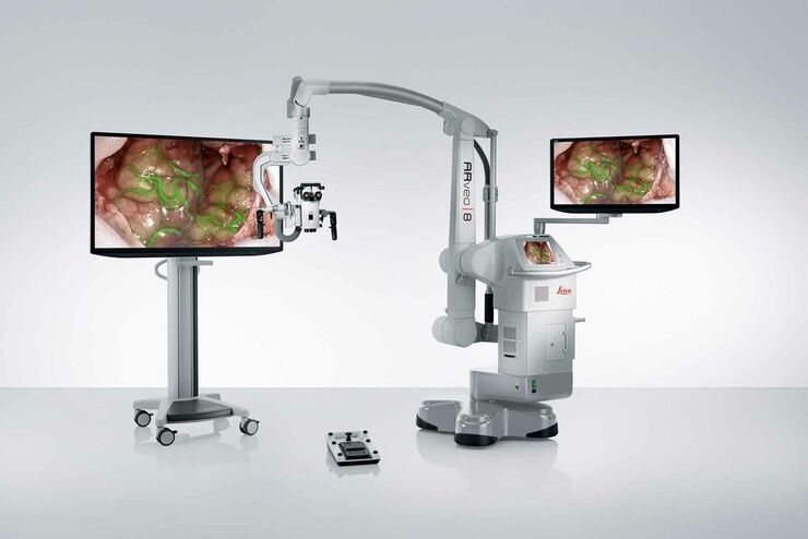
Next-generation digital visualization microscope from Leica Microsystems
News Archive
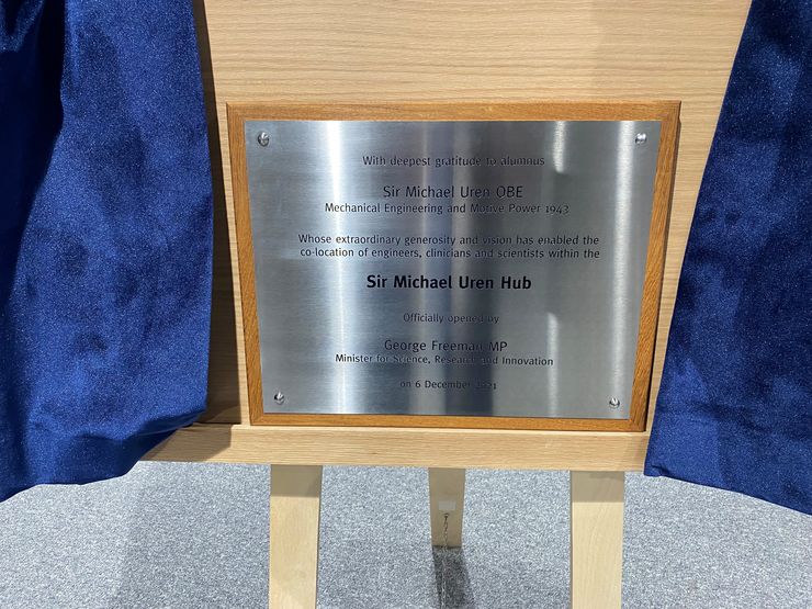
Leica Microsystems supports innovation through partnership agreement
4K camera in ENT microscope supports patient communication
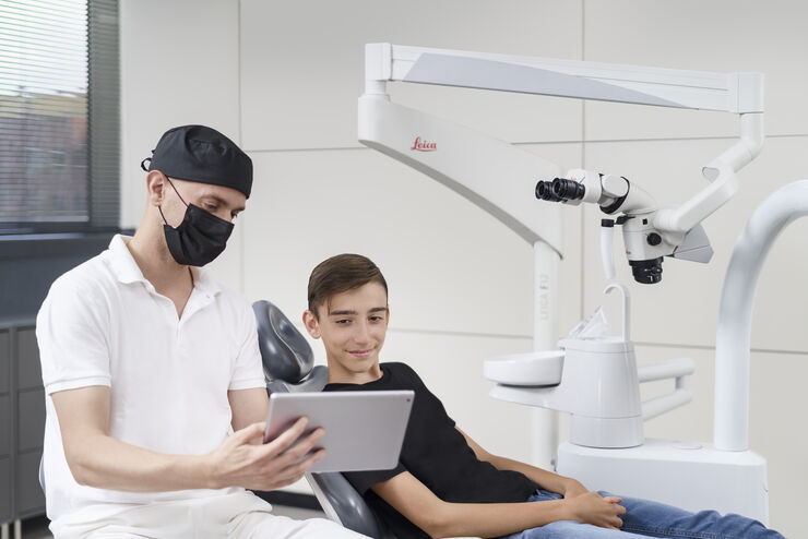
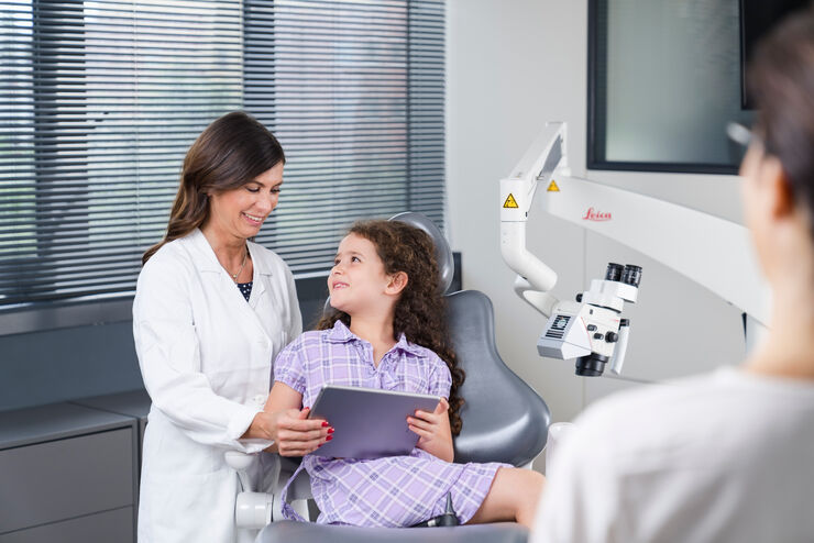
Dental microscope with new integrated 4K camera

Suitable for a wide range of brightfield and entry-level fluorescence quantitative imaging applications in life science and industry
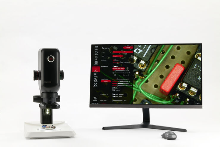
All-In-One Digital Inspection Solution from Leica Microsystems

Experience new functions with a free trial on the Aivia web platform

New solution promises to accelerate experimental success and improve process reliability as industry looks to faster drug discovery post-Covid

Wetzlar, Germany, March 16th 2021. Leica Microsystems, a world-leading designer and manufacturer of opto-digital imaging solutions in the field of…
19th November 2020 | Join Simona Paladino, from Naples University, in a live webcast with Nature to learn more about improvement of imaging techniques…
Join a Conversation about Structural Biology & Functional Imaging
Wednesday 16th September
London 16:00 | Berlin 17:00 | Dubai 19:00

Wetzlar and Berlin, May 2020. — Leica Microsystems is pleased to announce the donation of a THUNDER 3D Live Cell imaging system to the Institute of…
