
Especialidades médicas
Especialidades médicas
Explore una completa colección de recursos científicos y clínicos adaptados a los profesionales sanitarios, que incluye opiniones de colegas, estudios de casos clínicos y simposios. Diseñada para neurocirujanos, oftalmólogos y especialistas en cirugía plástica y reparadora, otorrinolaringología y odontología. Esta colección destaca los últimos avances en microscopía quirúrgica. Descubra cómo las tecnologías quirúrgicas de vanguardia, como la fluorescencia AR, la visualización 3D y las imágenes OCT intraoperatorias, permiten tomar decisiones con confianza y precisión en cirugías complejas.

Physiology Image Gallery
Physiology is about the processes and functions within a living organism. Research in physiology focuses on the activities and functions of an organism’s organs, tissues, or cells, including the…

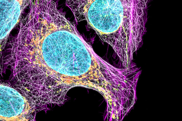
Live Cell Imaging Gallery
Live cell microscopy techniques are fundamental to get a better understanding of cellular and molecular function. Today, widefield microscopy is the most common technique used to visualize cell…

Tissue Image Gallery
Visual analysis of animal and human tissues is critical to understand complex diseases such as cancer or neurodegeneration. From basic immunohistochemistry to intravital imaging, confocal microscopy…
 and astrocytes (green) in a cortical spheroid derived from human induced pluripotent stem cells.")
Neuroscience Images
Neuroscience commonly uses microscopy to study the nervous system’s function and understand neurodegenerative diseases.

Multicolor Image Gallery
Fluorescence multicolor microscopy, which is one aspect of multiplex imaging, allows for the observation and analysis of multiple elements within the same sample – each tagged with a different…
Cell Biology Image Gallery
Cell biology studies the structure, function and behavior of cells, including cell metabolism, cell cycle, and cell signaling. Fluorescence microscopes are an integral part of a cell biologist…

Developmental Biology Image Gallery
Developmental biology explores the development of complex organisms from the embryo to adulthood to understand in detail the origins of disease. This category of the gallery shows images about…

Introduction to Widefield Microscopy
This article gives an introduction to widefield microscopy, one of the most basic and commonly used microscopy techniques. It also shows the basic differences between widefield and confocal…
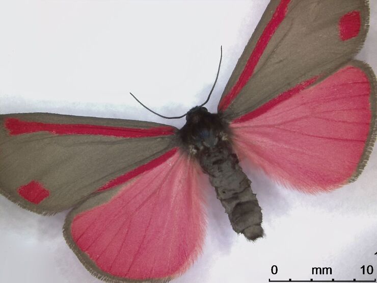
Life Science Imaging with DVM6 Digital Microscope
Digital microscopes can be a great help in life science applications such as the documentation in botany, entomology studies and crop science, or the digitization of museum collections. The image…
