Especialidades médicas
Especialidades médicas
Explore una completa colección de recursos científicos y clínicos adaptados a los profesionales sanitarios, que incluye opiniones de colegas, estudios de casos clínicos y simposios. Diseñada para neurocirujanos, oftalmólogos y especialistas en cirugía plástica y reparadora, otorrinolaringología y odontología. Esta colección destaca los últimos avances en microscopía quirúrgica. Descubra cómo las tecnologías quirúrgicas de vanguardia, como la fluorescencia AR, la visualización 3D y las imágenes OCT intraoperatorias, permiten tomar decisiones con confianza y precisión en cirugías complejas.
A New Method for Convenient and Efficient Multicolor Imaging
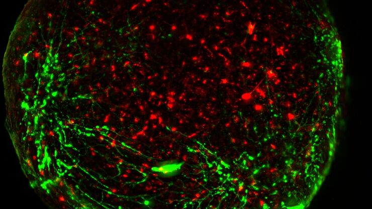
The technique combining hyperspectral unmixing and phasor analysis was developed to simplify the process of getting images from a sample labeled with multiple fluorophores. This aggregate method…
Considerations for Multiplex Live Cell Imaging
Simultaneous multicolor imaging for successful experiments: Live-cell imaging experiments are key to understand dynamic processes. They allow us to visually record cells in their living state, without…
How to Improve Live Cell Imaging with Coral Life
For live-cell CLEM applications, light microscopy imaging is a critical step for identifying the right cell in the right state at the right time. In this article, Leica experts share their insights on…
Microscopios de campo oscuro
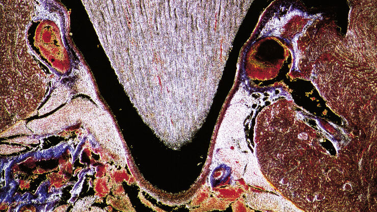
El método de contraste de campo oscuro aprovecha la difracción o dispersión de la luz de las estructuras de un espécimen biológico o las características no uniformes de una muestra de material.
Download The Guide to Live Cell Imaging
In life science research, live cell imaging is an indispensable tool to visualize cells in a state as in vivo as possible. This E-book reviews a wide range of important considerations to take to…
The Power of Pairing Adaptive Deconvolution with Computational Clearing
Learn how deconvolution allows you to overcome losses in image resolution and contrast in widefield fluorescence microscopy due to the wave nature of light and the diffraction of light by optical…
Studying Pulmonary Fibrosis
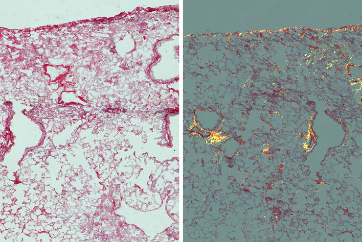
The results shown in this article demonstrate that fibrotic and non-fibrotic regions of collagen present in mouse lung tissue can be distinguished better with polarized light compared to brightfield.…
Image Gallery: THUNDER Imager
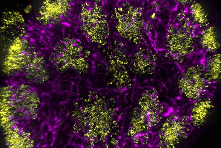
To help you answer important scientific questions, THUNDER Imagers eliminate the out-of-focus blur that clouds the view of thick samples when using camera-based fluorescence microscopes. They achieve…
From Organs to Tissues to Cells: Analyzing 3D Specimens with Widefield Microscopy
Obtaining high-quality data and images from thick 3D samples is challenging using traditional widefield microscopy because of the contribution of out-of-focus light. In this webinar, Falco Krüger…


