
Filter articles
タグ
Loading...
 in 14 x 18 tiles. Lifetime gives an additional contrast that allows to differentiate different structures in histological stainings.")
A Guide to Fluorescence Lifetime Imaging Microscopy (FLIM)
The fluorescence lifetime is a measure of how long a fluorophore remains on average in its excited state before returning to the ground state by emitting a fluorescence photon.
Loading...

Live-Cell Imaging Techniques
The understanding of complex and/or fast cellular dynamics is an important step for exploring biological processes. Therefore, today’s life science research is increasingly focused on dynamic…
Loading...

Step by Step Guide for FRAP Experiments
Fluorescence Recovery After Photobleaching (FRAP) has been considered the most widely applied method for observing translational diffusion processes of macromolecules. The resulting information can be…
Loading...
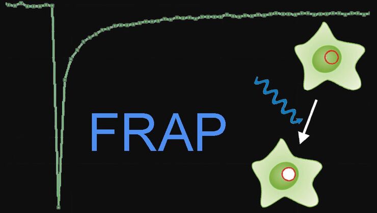
Fluorescence Recovery after Photobleaching (FRAP) and its Offspring
FRAP (Fluorescence recovery after photobleaching) can be used to study cellular protein dynamics: For visualization the protein of interest is fused to a fluorescent protein or a fluorescent dye. A…
