Background image

Microscopy, Imaging and Analysis Solutions from Leica Microsystems

We shape the future by empowering users of our breakthrough optical and digital solutions to create a better, healthier world. For more than 175 years, we have been revealing the invisible with our market-leading technology in life science research, industry, medicine, forensics and education.

Revealing the Invisible for a Better and Healthier World.

Main Areas We Serve

Medical Specialties

With a portfolio covering microsurgery disciplines including ophthalmology, neurosurgery, otolaryngology (ENT), plastic reconstructive surgery and dentistry, there is a surgical microscope from Leica to meet your needs.

Life Science Research

Leica Microsystems’ life science research microscopes support the imaging needs of the scientific community with advanced innovation and technical expertise for the visualization, measurement and analysis of microstructures.

Industrial Microscopy

Maximizing uptime and achieving targets efficiently help your bottom line. Leica Microscope solutions can give you insights into the smallest sample details as well as analyze, document, and report results fast and reliably.

Environmental stewardship: Plant growing

Our Commitment to Sustainability

At Leica Microsystems, we believe in the power of science and innovation to build a more sustainable future. Our sustainability strategy is built on three core pillars: Innovating for a Better World, Caring for Our People, and Protecting Our Planet. By integrating these principles into our business practices, we drive progress and create a positive impact on society and the environment.

Read More

Introducing Online Shopping

Introducing Online Shopping

Find the Perfect Solution for Your Application Needs.

Access our extensive product catalog and enjoy a seamless online shopping experience. Explore and purchase pre-configured bundles in our Leica Microsystems shop.

Shop is not offered in every market.

Read More

Advanced Imaging – DMi8 Inverted Microscope Platform

Streamline your complex workflows and generate high quality data.

Streamline your complex microscopy workflows with a platform that enables you to generate data with solutions that you can tailor to your research requirements and budget.

Read More

Look beyond - MyVeo Headset

MyVeo All-In-One Surgical Visualization Headset

Stay focused, work comfortably, and enhance team collaboration in the OR.

Have you imagined what the future of surgery will look like? Reimagine how you see, how you move, and how you collaborate during surgery – MyVeo takes you beyond what you’ve imagined.

Read More

Ivesta 3 Greenough Stereo Microscopes

Ivesta 3 Greenough Stereo Microscopes

What if you could inspect with increased efficiency and work in a consistent way?

For manufacturers or suppliers, increasing efficiency for inspection is a priority. Optimize your visual inspection and rework while achieving reliable, consistent results.

Read More

Aivia AI Image Analysis Software

Access the future of AI microscopy

Aivia is a complete 2-to-5D image visualization, analysis and interpretation platform designed for the reliable processing and reconstruction of highly complex images.

Read More

Environmental stewardship: Plant growing
Introducing Online Shopping
Look beyond - MyVeo Headset
Ivesta 3 Greenough Stereo Microscopes

Our Latest Articles

Read our latest articles

The knowledge portal of Leica Microsystems offers scientific research and teaching material on the subjects of microscopy. The content is designed to support beginners, experienced practitioners and scientists alike in their everyday work and experiments.

More Articles

3D rendering of a mouse beta cell with mitochondria (blue)
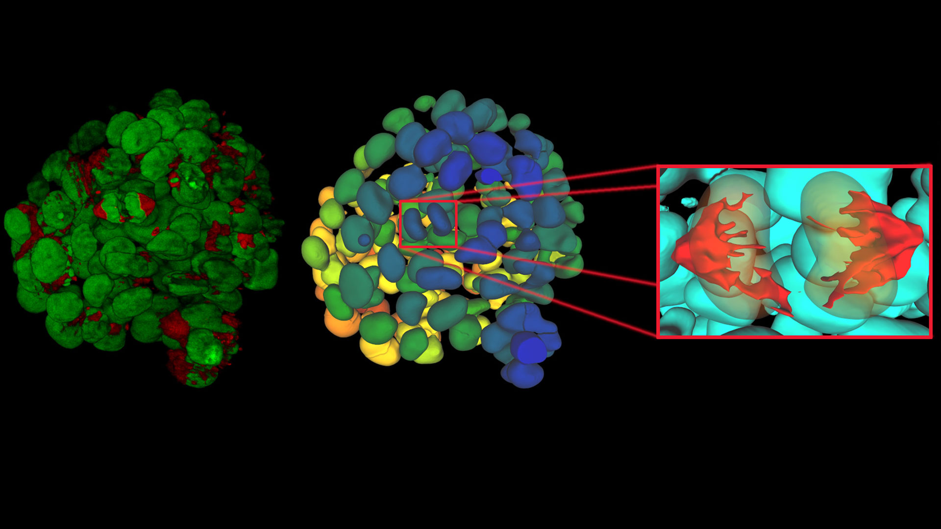
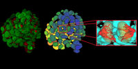
, insulin SGs (orange), microtubules (red), nucleus (yellow), and plasma membrane (transparent).

High-Pressure Freezing Protocols for Ultrastructural 3D EM

High pressure freezing (HPF) can help preserve hydrated cells and tissues close to their biological state at the moment of immobilization, supporting more reliable ultrastructural interpretation than…
Claudia Mayrhofer ZFE Graz

Ultramicrotome UC Enuity in Practice: Stable 15 nm Sections at ZFE

After using the UCT and UC6 ultramicrotomes, Claudia Mayrhofer calls UC Enuity a leap in stability—so robust that vibrations and temperature shifts don’t spoil sections, even with multiple users. Auto…
Image of glass with primary corundum having numerous inclusions. It was taken with a microscope using polarization contrast.

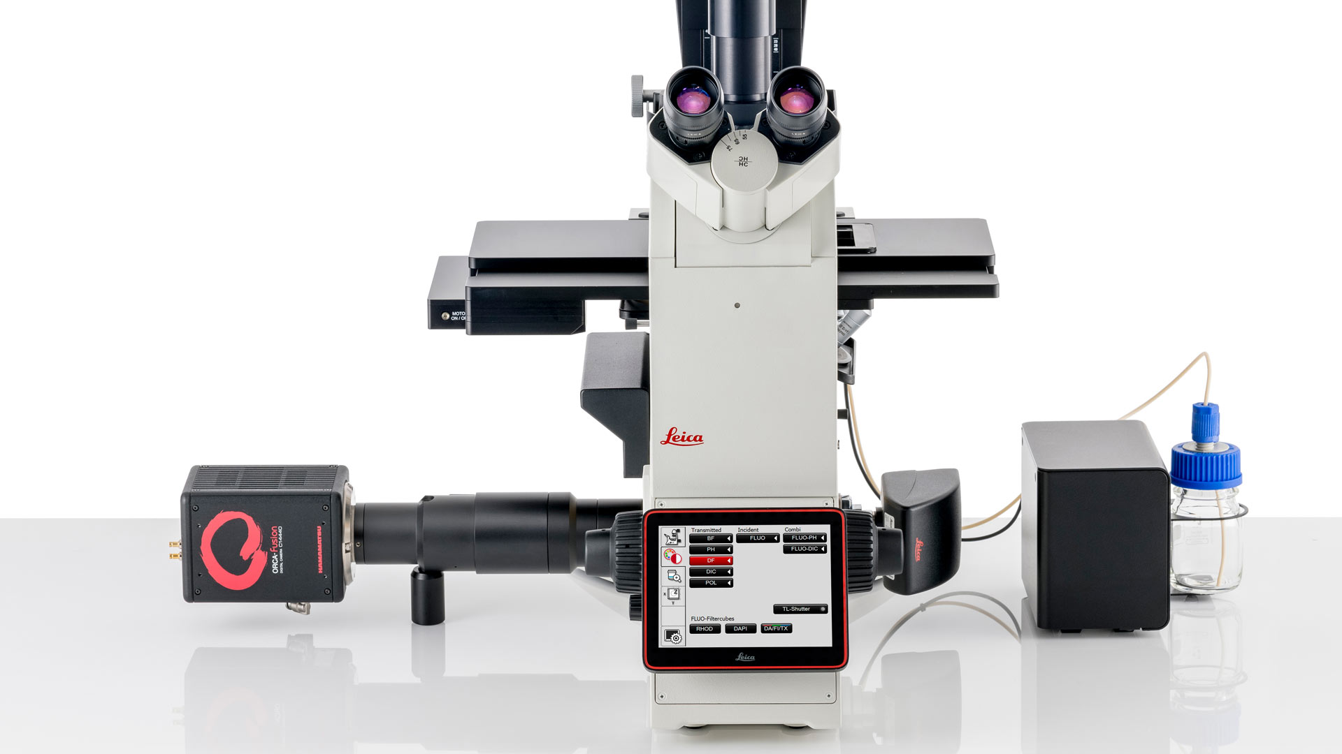
Ensuring Glass Quality with the Polarization Microscopy Advantage

Glass is one of the oldest materials known. Today, it is used for many applications, e.g., optical instruments, windows, doors, solar panels, containers for food, beverages, and medicine, so strict…
OCSS Cataract Surgery Symposium

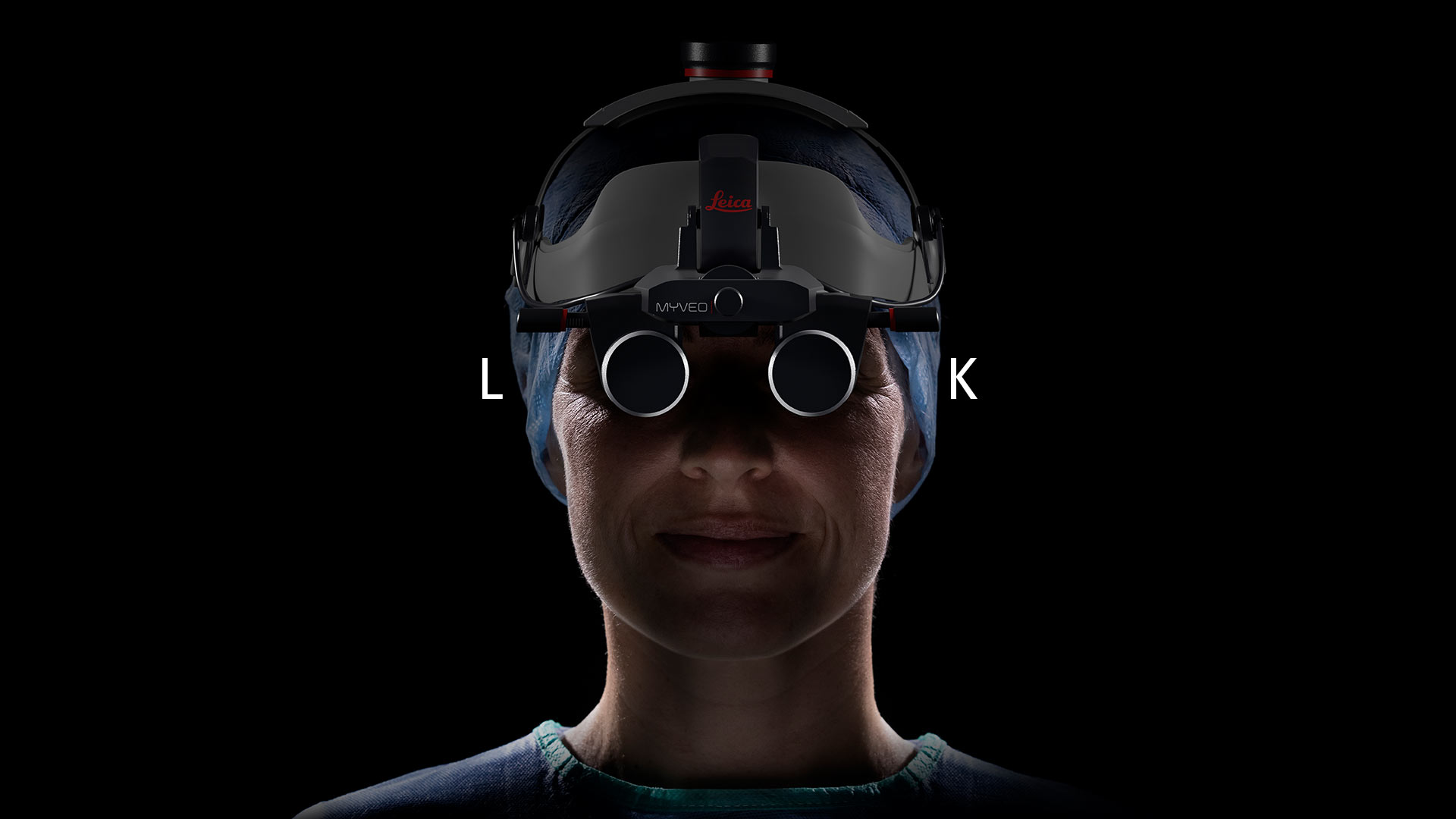
Expert Techniques for Superior Visualization in Cataract Surgery

Join renowned ophthalmic surgeons, Dr. Hussein Almuhtaseb and Mr. Simon Madge, as they share their clinical expertise and real-world surgical strategies during the 2025 Online Cataract Surgery…
LMD ionizer before & after cut.

Eliminating Electrostatic Interference in Laser Microdissection

Electrostatic charge in laser microdissection (LMD) causes two critical failures: samples stick to charged surfaces and are lost, or samples fly into adjacent wells and cause cross-contamination. We…

History, Developments and Trends of Microscopy in Cancer Research

Cancer is a global disease, with 18 million new cases diagnosed and 10 million cancer-related deaths worldwide in 2020. This burden is set to increase, with a projected increase in cases of ~55% by…
Mouse fibroblasts where F-actin is labelled with FITC (green), tubulin with Cy5 (red), and nuclei with DAPI (blue). Image courtesy of Dr. Günter Giese, Max Planck Institute for Medical Research, Heidelberg, Germany.

Overview of Fluorescent Dyes in terms of Applications and Properties

An introduction to commonly used fluorescent dyes and an overview of their characteristics are given in this article. Fluorescence microscopy is used for the study of specific cellular components with…

Researchers Insights: Microscopy in Cancer Research

Discover how imaging techniques are driving cancer research forward. In this issue, we present comprehensive multimodal studies using microscopy, as well as new directions in intraoperative cancer…
Leica Microsystems Life Science Product

Predictive Service Prevents Downtime in Ghent

At the VIB BioImaging Core in Ghent, Belgium, researchers depend on Leica’s Stellaris 8 confocal microscope to explore the frontiers of biomedical science. When Leica’s RemoteCare system detected a…
Zebrafish heart, DAPI (nuclei, blue), Tropomyosin (cardiomyocytes, red) and GFP (primordial cardiac layer, green). Courtesy of Anna Jazwinska, University of Fribourg, Switzerland.

A Guide to Fluorescence Microscopy

Fluorescence microscopy uses the ability of fluorophores, dyes, or fluorescent proteins to emit light of a specific wavelength after being excited with light of a shorter wavelength. Biomolecules can…
Petri dishes lab environment

ATTO-TEC Consumables

ATTO-TEC dyes have become a benchmark for fluorescence microscopy imaging, offering a highly differentiated panel. Their brightness and photostability make them the reagents of choice for demanding applications.

Learn more

10-14 June 2023, Heidelberg, Germany – the course focusing on fluorescence lifetime-based readouts organized with the EMBL Imaging Centre gathered…

EM ICE demonstrated during the course by Andreia Pinto

The EMBO (European Molecular Biology Organization) practical course took place between the 12th and the 17th of February 2023. It covered all steps of…

Marko Lampe (Scientific Officer at the Advanced Light Microscopy Facility, EMBL) setting up the STELLARIS 8 STED for the virtual course

Jointly organized by Leica Microsystems and EMBL, this virtual course held between the 8th and 15th of July 2022, provided attendees with deeper…

EMBL IC (courtesy of EMBL)

The EMBL Imaging Centre was inaugurated on the 30th of June 2022, welcoming representatives from politics, industry and research. The center was…

Leica booth before official opening of the event

The EMBL in Heidelberg hosted its first Scientific Symposium at the EMBL Imaging Centre on the 30th of May 2022, featuring talks about leading edge…

Scroll to top