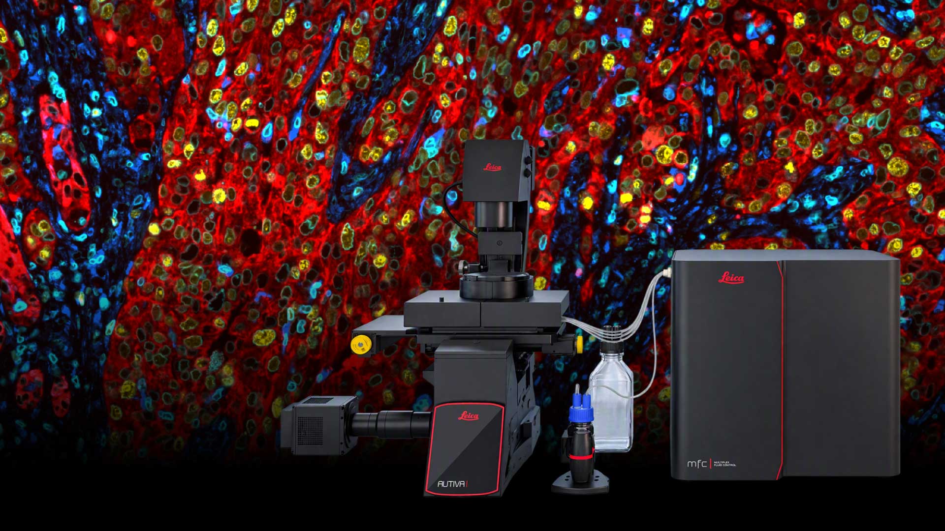
Autiva Spatial Phenotyper
Spatial insights start with an image. Autiva Spatial Phenotyper combines Leica Microsystems’ imaging expertise with automated staining, an expanding reagent ecosystem, and AI-powered image analysis to create a comprehensive workflow that captures richer spatial data, reveals meaningful cellular relationships, and provides a deeper understanding of complex biology in every sample.
Productivity where it counts
Spatial biology studies continue to grow in size and complexity. The Autiva Spatial Phenotyper helps laboratories scale their research without scaling complexity.
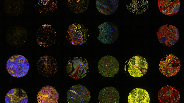
By integrating cyclic staining and imaging into a highly automated workflow, the system enables efficient analysis of tissue microarrays (TMAs), whole tissue sections, and larger study cohorts. Researchers can process more samples with less hands-on time, accelerating data generation and discovery.
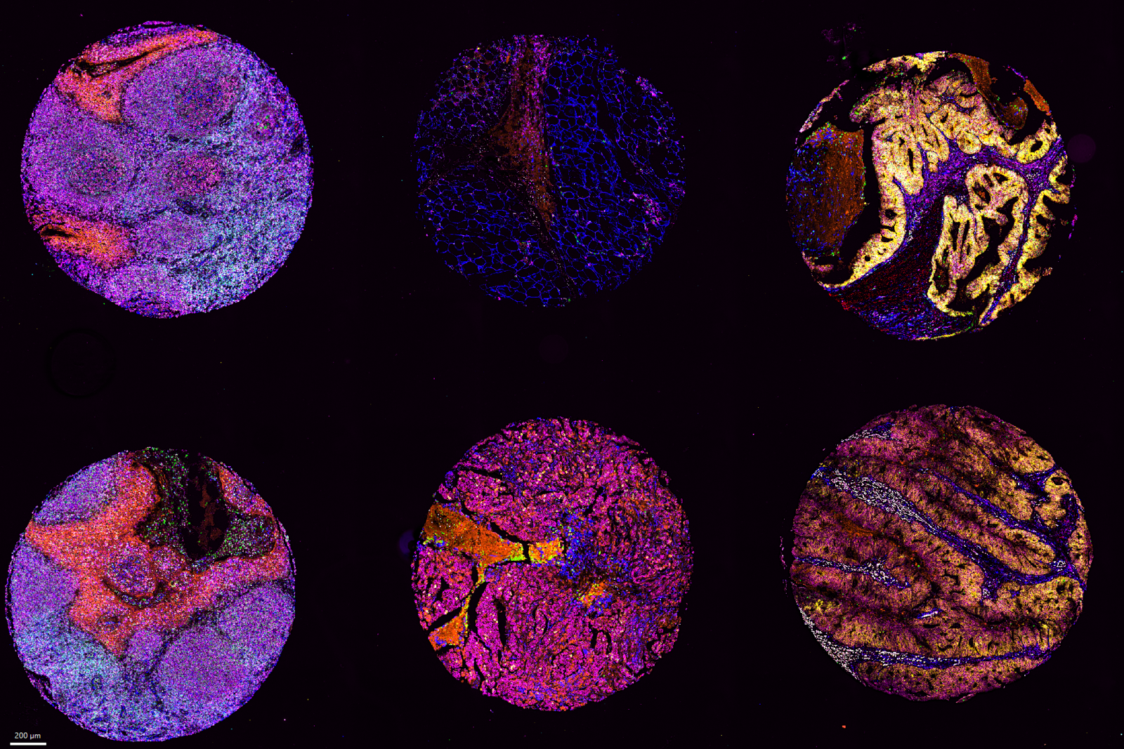
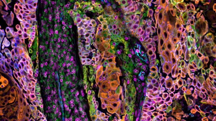
Credit: Federica Sella and Evelyn Lattmann (USZ Zurich, CH, Mitch Levesque Group)
Move from sample to spatial insight faster
Go from staining to quantitative spatial insights through a walk-away workflow that automates both staining and imaging. Process up to four slides in parallel while reducing manual intervention and workflow bottlenecks.
- Deliver industry-leading throughput with up to 3× higher sample capacity
- Process 24 biomarkers across four slides in less than 18 hours

Capture more biology per experiment
Simultaneous multi-channel imaging reduces the number of imaging cycles required, enabling researchers to generate highly multiplexed datasets more efficiently, preserve sample quality, and accelerate time to results.
- Capture seven fluorescence channels per cycle and generate virtual H&E images with every dataset
- Image up to 614 mm² of tissue per slide, supporting comprehensive analysis of large tissue sections in a single run
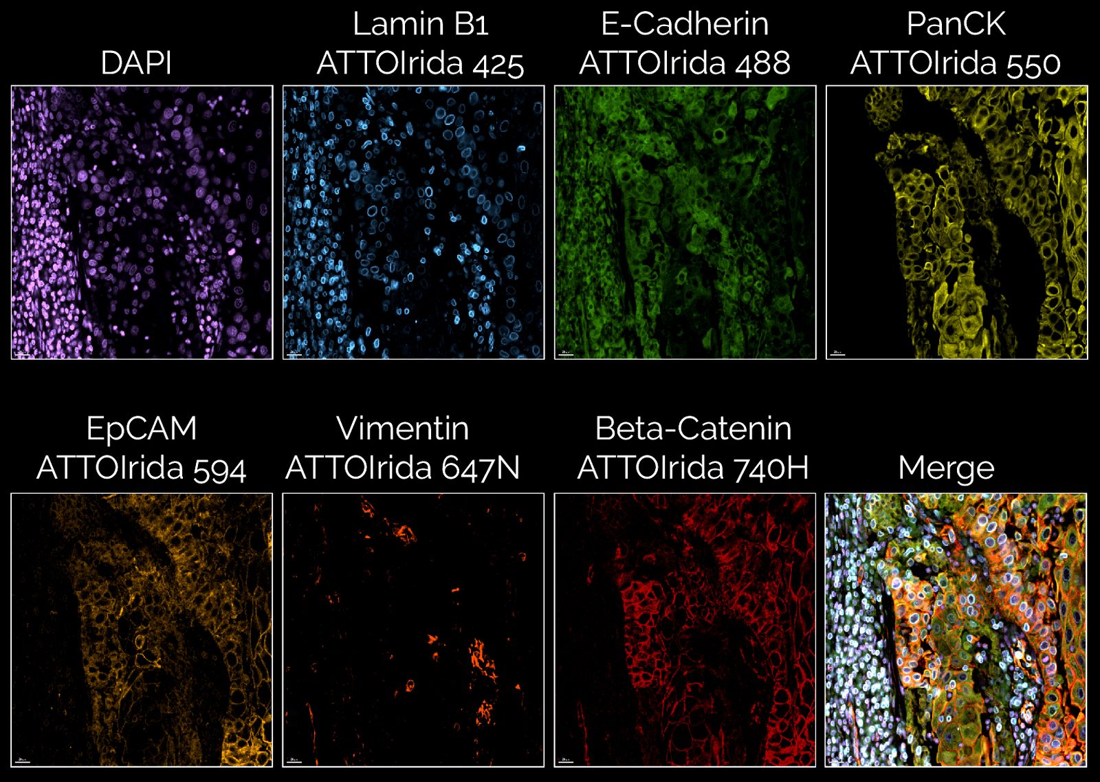
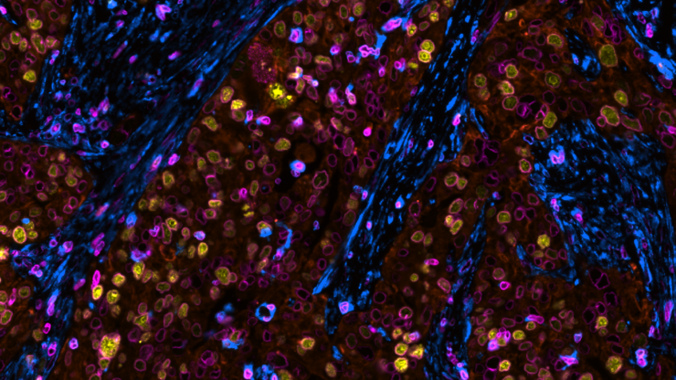
Spatial insights start with an image.
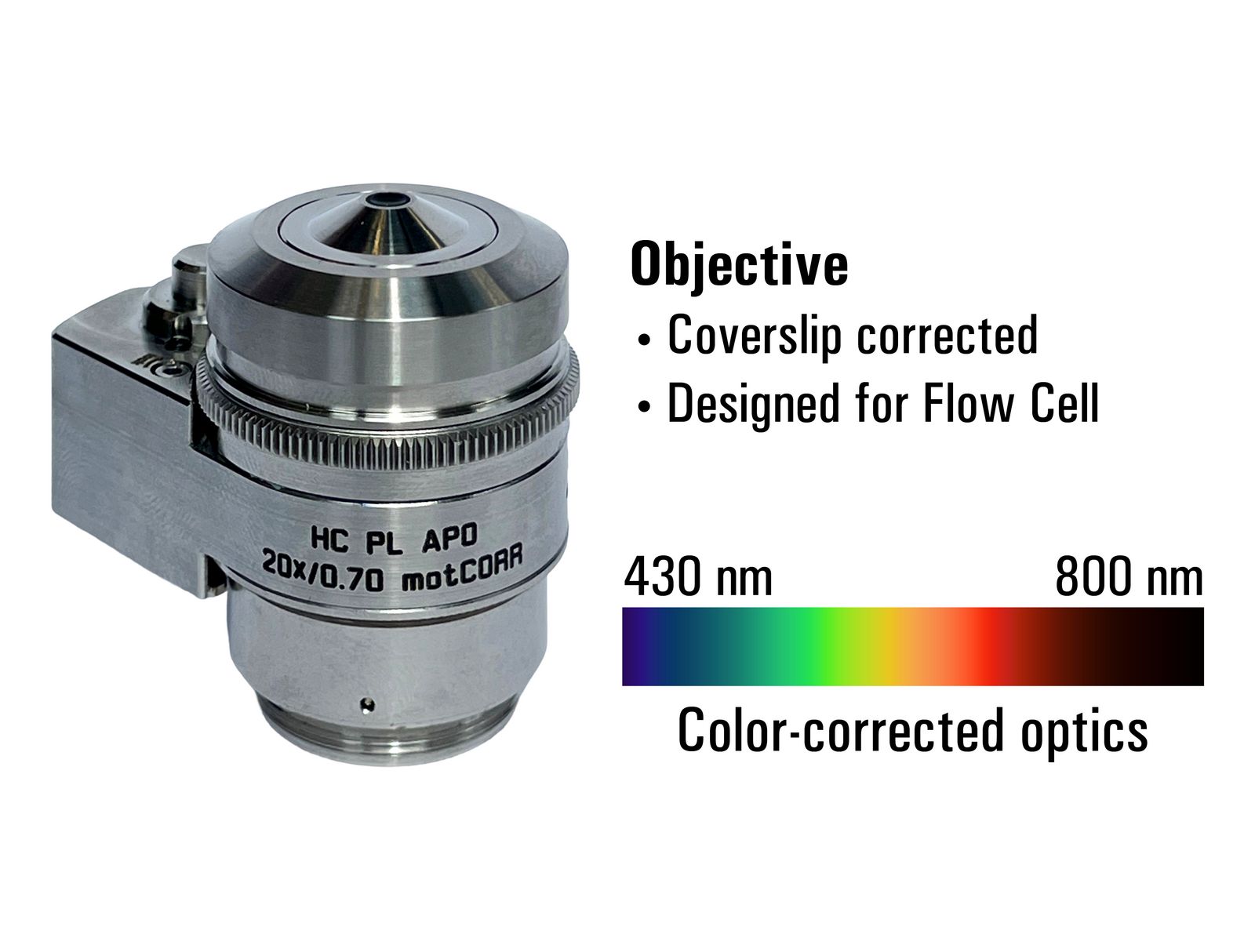
The Autiva Spatial Phenotyper combines Leica Microsystems' imaging heritage with a custom objective engineered specifically for spatial biology workflows. The imaging system delivers the image quality needed for accurate segmentation, phenotyping, and spatial analysis.
- Coverslip-corrected and optimized for Flow Cell imaging
- Color-corrected from 430 to 800 nm to support multiplex imaging across a broad fluorescence spectrum
See biology with greater confidence
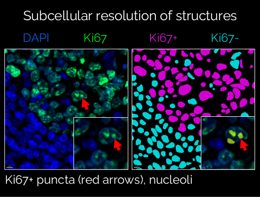
High-resolution imaging resolves cellular and subcellular features across large tissue regions, enabling researchers to visualize fine morphological details and complex tissue architecture.
The dedicated optical path is optimized for spatial biology workflows, supporting accurate cell segmentation, cell identification, phenotyping, and quantitative spatial analysis. By capturing small biological structures with clarity, researchers can more effectively characterize spatial relationships and biological heterogeneity within every sample.
Built on proven high-plex expertise
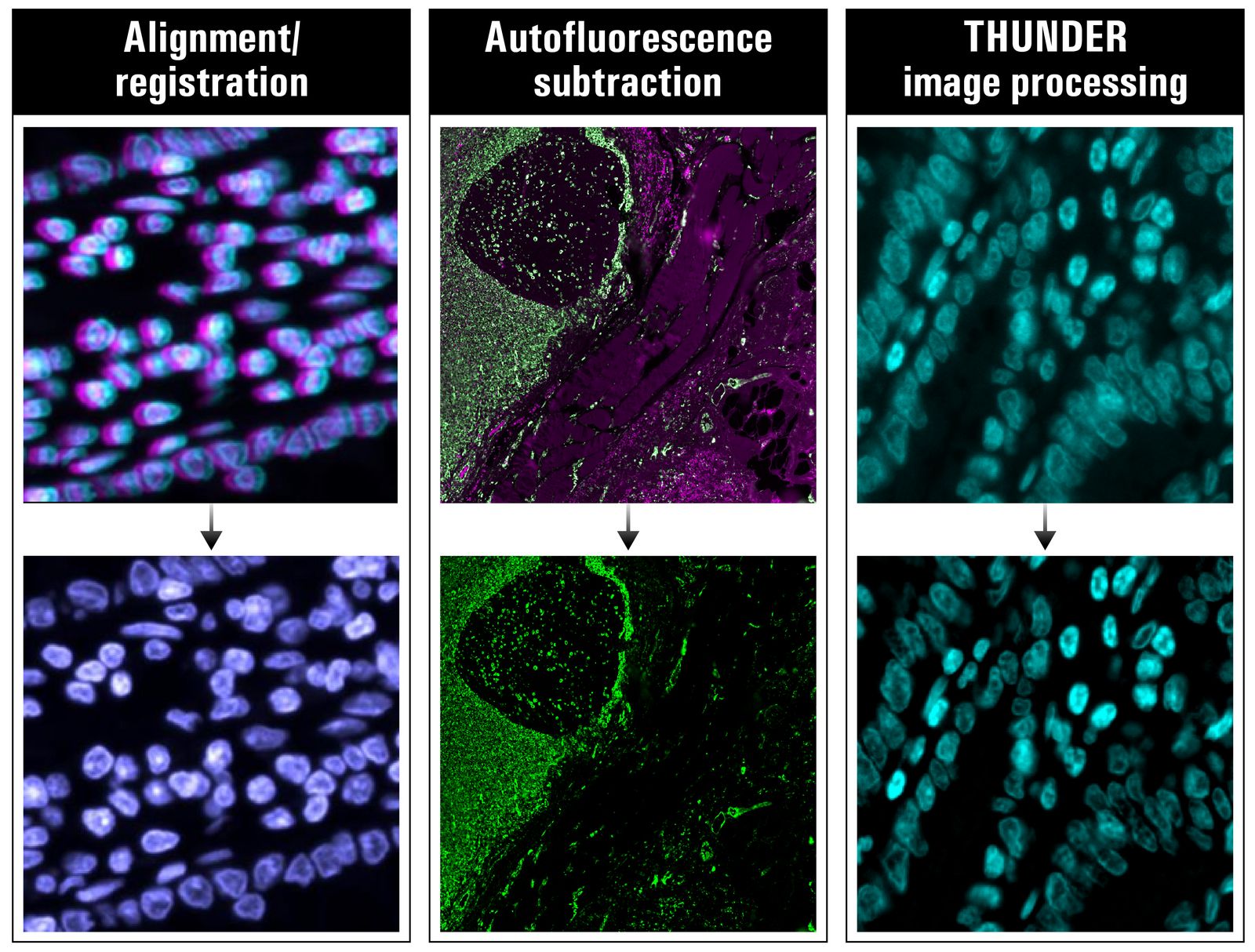
The Autiva Spatial Phenotyper combines more than a decade of high-plex imaging experience from the Cell DIVE platform with Leica Microsystems' long heritage in imaging. Advanced calibration and image correction workflows help generate high-quality, analysis-ready images for spatial biology.
- Automated registration, distortion correction, flat-field correction, and autofluorescence removal
- THUNDER Computational Clearing enhances contrast and reduces haze to generate high contrast images
Flexibility where it counts
Start with the capabilities you need today. Grow as your science evolves.
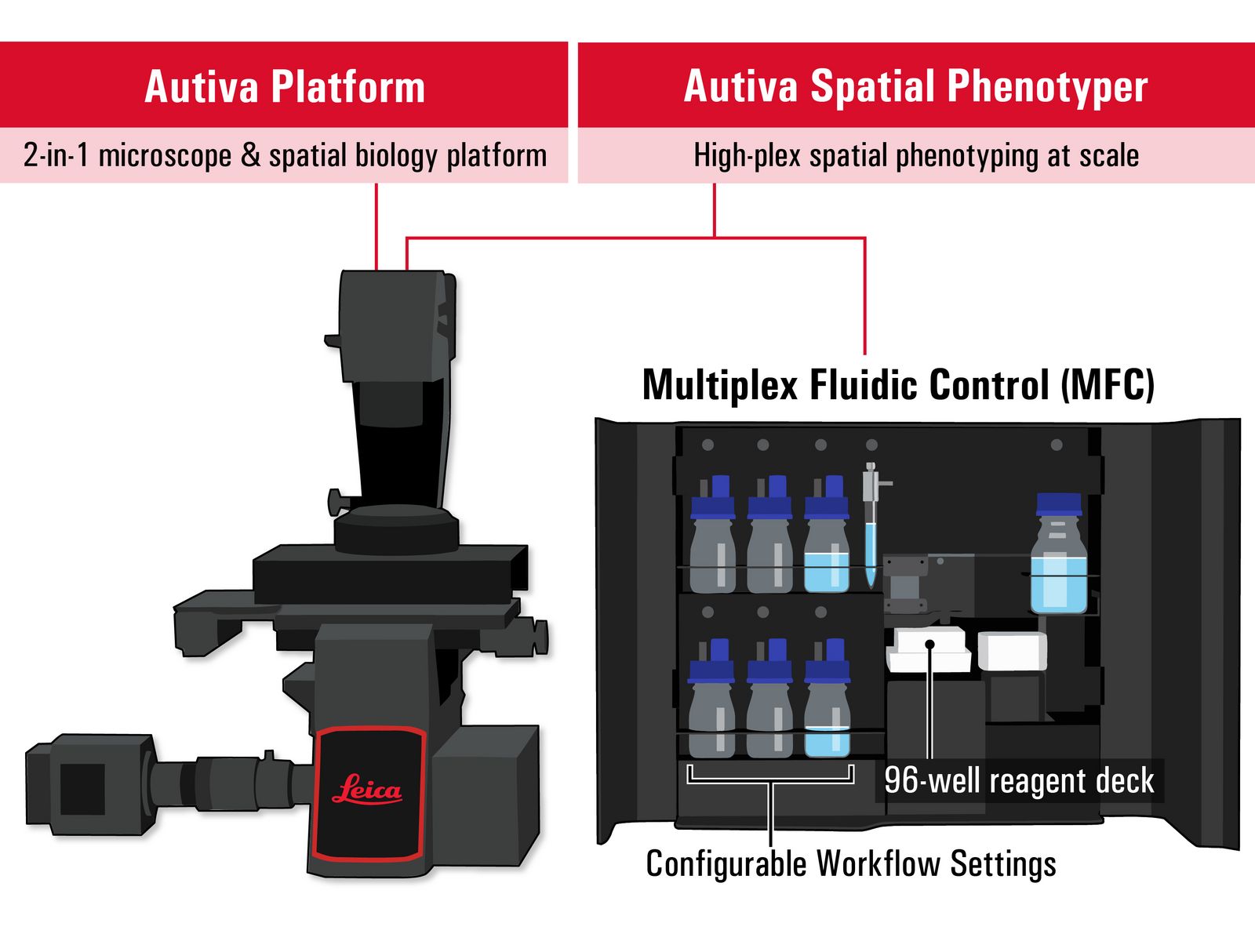
The Autiva Platform functions as both a microscope and an open spatial biology platform. Configurable workflow settings, a 96-well reagent deck, and flexible automation architecture support a broad range of current and future spatial biology workflows.
Researchers can expand into new applications over time without being locked into one approach or replacing their system.
An ecosystem designed to expand
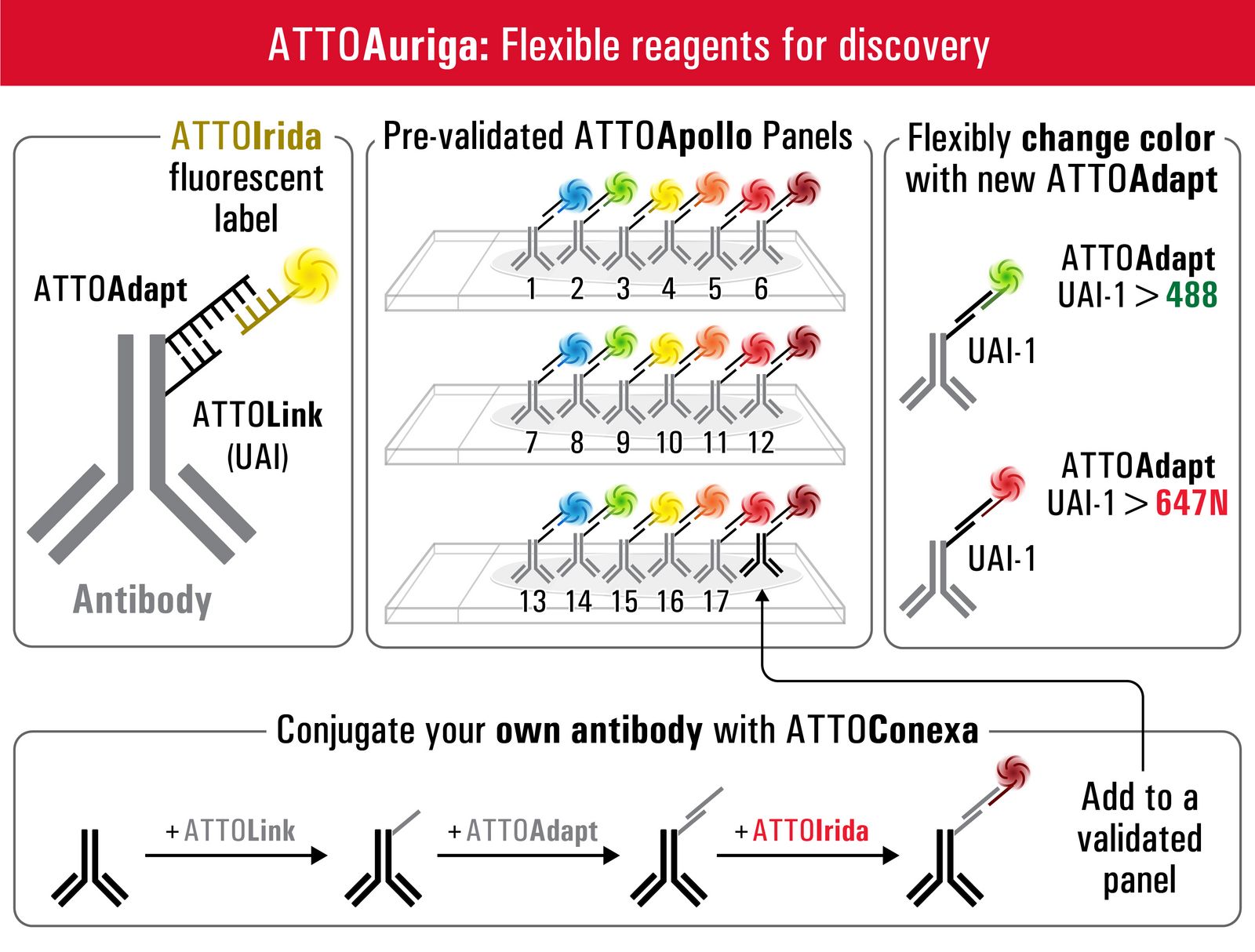
Grow your multiplex experiments with a reagent ecosystem designed to expand with your research. ATTOAuriga helps researchers start with validated ATTOApollo panels, flexibly reassign fluorophores using ATTOAdapt reagents, and add custom markers with ATTOConexa as research questions evolve.
The result is a standardized yet flexible approach that allows researchers to build upon validated panel content and incorporate custom markers as research needs change.
Transform data into discovery
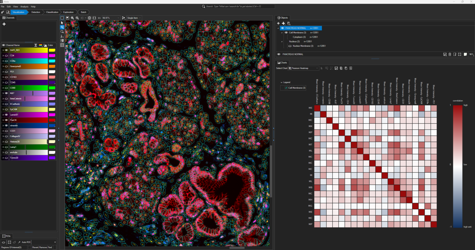
Turn complex spatial images into actionable biological insight with Aivia, AI-powered image analysis software designed for spatial biology. Cell membrane segmentation converts images into individual cellular objects that can be quantified, classified, and explored in context.
Researchers can uncover patterns through heat maps, correlation plots, clustering, and phenotyping analyses, with flexible licensing options to fit their laboratory needs.