GLOW800 Augmented Reality Fluorescence Application
Vascular Surgery. Augmented
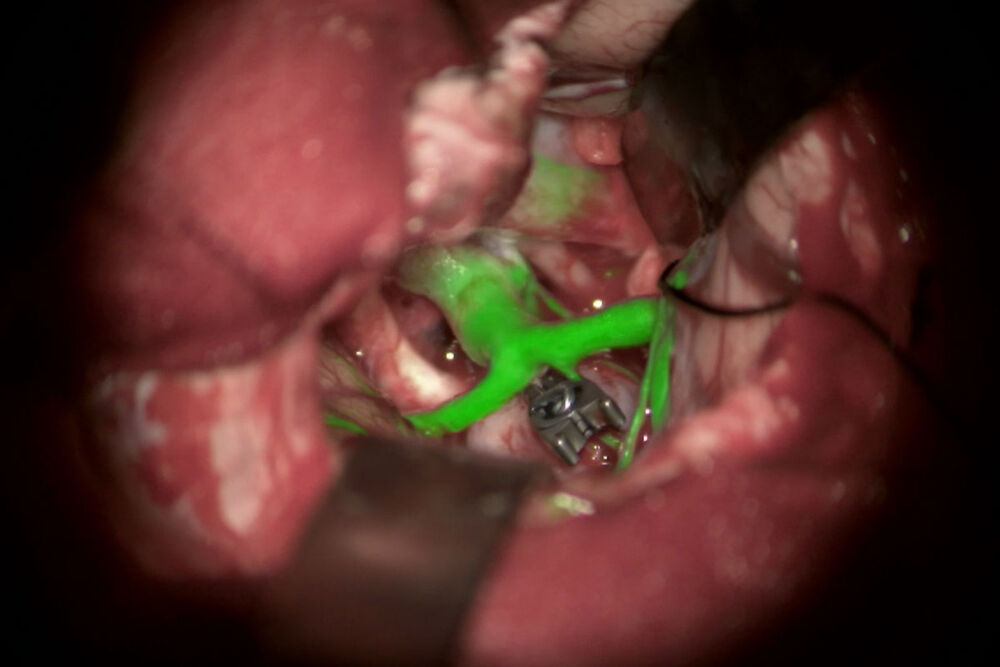
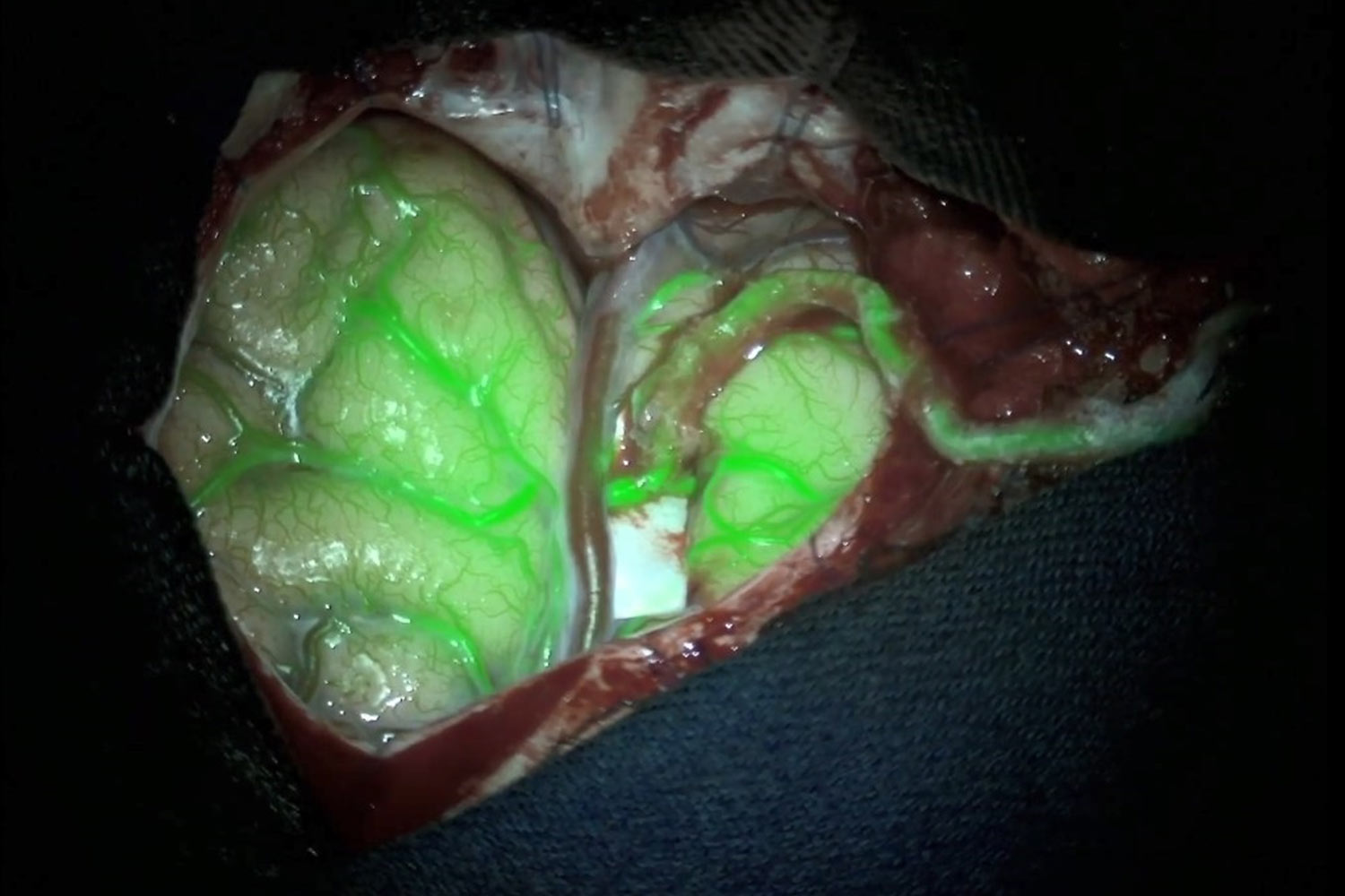
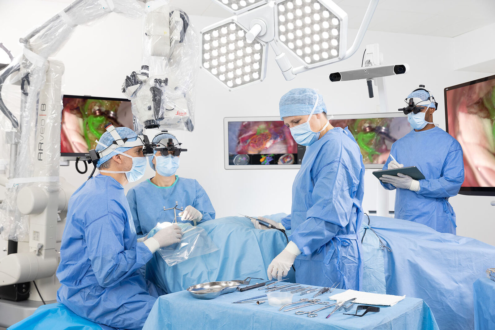
Whether clipping an aneurysm, removing an AVM, performing a microvascular decompression or a bypass, your ability to clearly view and assess cerebral anatomy and blood flow is critical for an optimal surgical outcome. With the GLOW800 augmented reality (AR) fluorescence application and ICG, you can observe cerebral anatomy in natural colour, augmented by real-time vascular flow, with 3D visualization.
Experience the benefits of one augmented view during vascular neurosurgery for enhanced confidence in your surgical intervention.
One complete picture of the cerebro- vascular region
- One augmented, real-time view means no more need to recall and try to reconcile the black-&-white blood flow video with the natural anatomical view
- Crisp delineation helps you limit potential compromise or obstruction of surrounding perforators and small vessels
- Digitally enhanced 3D depth perception and no dark periphery support clear spatial orientation to aid manipulation of vessels
Images courtesy of Cleopatra Charalampaki, MD, PhD, Professor of Neurosurgery, Department of Neurosurgery, Cologne Medical Center, Germany.


Advantages of GLOW800 AR compared to NIR imaging
Compare the visualization of cerebrovascular flow with the GLOW800 AR fluorescence application to traditional near infrared (NIR) imaging by watching this video.
View blood flow without interrupting workflow
No more interrupting surgery to switch between the natural microscope image and a flat black-&-white, near-infrared video. No more mental gymnastics to recall and reconcile the different views.
We are now really closer to what we believe is an augmented reality
Professor Raphael Guzman about GLOW800
Benefits for your surgery
Visualization with the GLOW800 AR application supports each step of your vascular neurosurgery procedure and also supports post-operative imaging.
For example during aneurysm clipping, it helps you:
- Confidently apply an aneurysm clip
- Assess clip placement and aneurysm occlusion
- Check if all branches proximal and distal to the clipped aneurysm are perfused and whether there is orthograde filling of the blood vessels
- Confirm the clip has not caused any compromise of surrounding blood vessels, such as kinking or partial obstruction

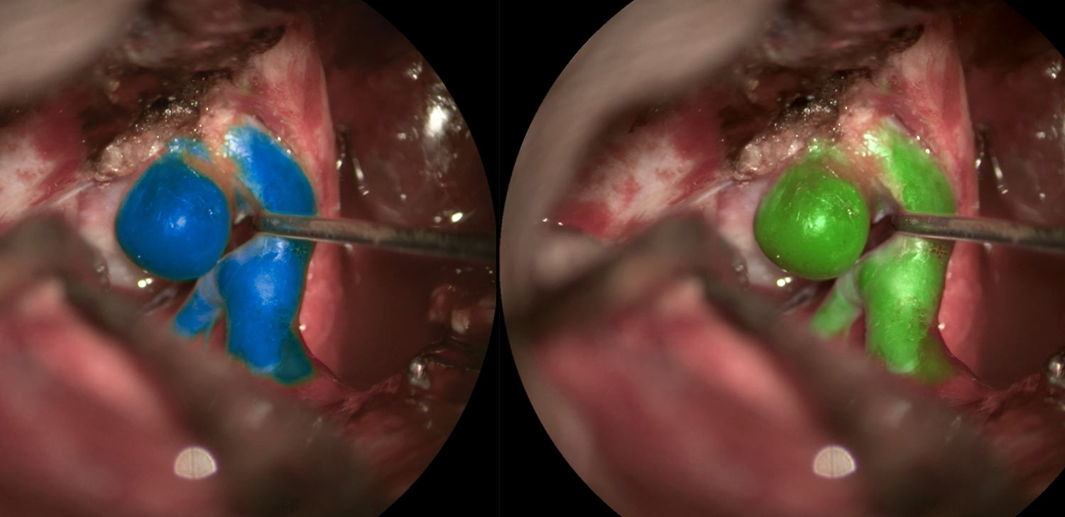
Choose your color!
Select a pseudo color according to your preference and for optimal contrast of the tissue.
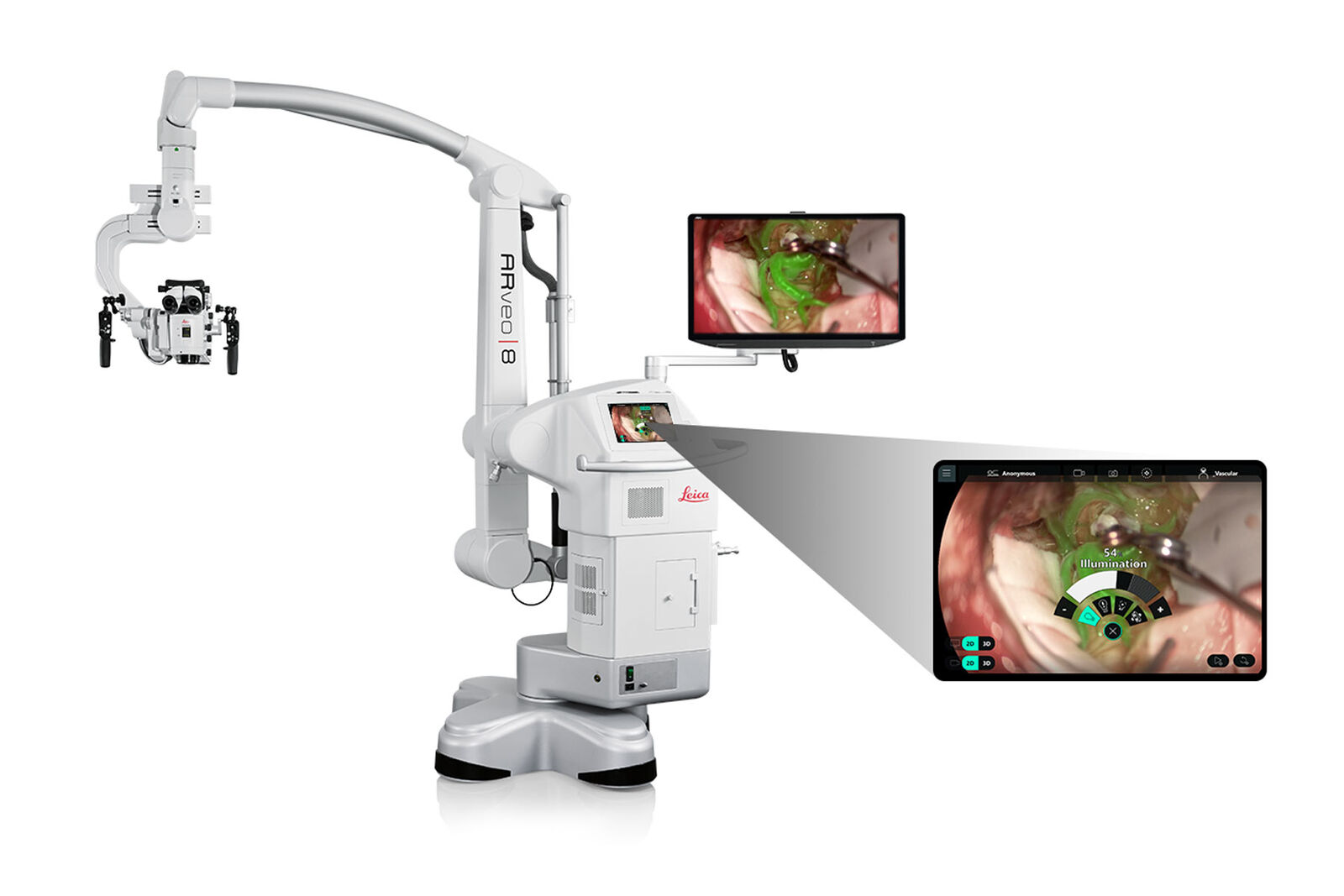
See what matters with flexible image settings
The GLOW800 application provides intuitive image parameters that allow you to customize the visualization of blood flow and tissue perfusion.
You can adjust the intensity, brightness, and threshold of fluorescence in the GLOW800 application to get to the image you need.
Experience optimal imaging in AVM, bypass, and aneurysm surgery, as well as for other surgical procedures like plastic reconstructive surgery.
Experience GLOW800 in highresolution 3D
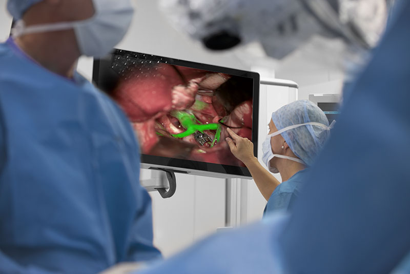
Let all members of your team participate in a shared real-time GLOW800 3D visualization via 3D monitor or with up to three MyVeo headsets.
GLOW800 allows for seamless integration into your procedure workflow, thanks to its compatibility with the ARveo 8* digital visualization microscope.
*Evolved ARveo 8 and ARveo 8x microscopes
Set up and record with ease
Display the GLOW800 application on a HD or 4K 3D monitor for the whole team. Then easily capture videos in 2D or 3D quality for later review and teaching outside the OR.
- One-touch activation of recording via the microscope handle or main screen of the ARveo 8 graphical user interface (GUI)
- Playback and review recordings easily utilizing the microscope handles, the footswitch or the quick menu of the ARveo 8 GUI.
2D visualization is only for observation, not for Heads-up display surgery.
GLOW AR Platform: The technology behind GLOW800
GLOW800 AR is the first clinical application of the GLOW AR platform based on digital spectral detection technology from Leica Microsystems.
The sophisticated imaging sensors and algorithms of GLOW AR acquire, optimize, and combine multiple spectral bands of light. The result is natural coloring of tissue anatomy and accurate representation of fluorescence intensity.