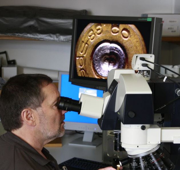

Ballistics: Fired ammunition and gunshot residue under the comparison macroscope
In the laboratory, the forensic experts subject the evidence they have collected to closer scrutiny. A large part of their work concerns ballistics – i.e. the examination of firearms and ammunition. If the perpetrator has used a firearm, one or more projectiles and bullet cases can usually be found at the scene of the crime. When the cartridge is fired, the case is first pulled back in the weapon and subsequently ejected. The next cartridge then moves into the barrel from the magazine. The ammunition collected at the crime scene is examined in more detail in the laboratory using optical magnification instruments, sometimes even special comparison macroscopes.
Comparison macroscopes, which are typically used for magnifications up to 100x, have a so-called comparison bridge that unites two complete light paths to form one combined image. The forensic analysts can choose between a split-image comparison, a comparison of the two full images or a superimposed version of the two images. Mostly, they choose the split-image option, which, for instance, enables side by side comparison of the projectile found at the crime scene with a test projectile fired in the laboratory. To have a projectile for comparison in the first place, the investigators first have to find the potential crime weapon. It may be registered in a database or, more probably, discovered when the potential perpetrator’s home is searched.


Test firing into the water tank
Once in possession of a potential crime weapon, the forensic examiners fire it in a water tank or similar bullet retrieval system. This effectively prevents the projectile from being deformed and being more difficult to evaluate later. The two projectiles – the one from the crime scene and the one from the comparison weapon – are then viewed with the comparison instrument. In the split-image view there is a thin black dividing line, at first usually in the center between the two image halves. However, this line can be moved sideways to show more of the left or the right image. By moving the dividing line horizontally, the examiner can see whether a trace from one object is continued in the next. The barrel of a gun, for example, is not straight, but always twisted. Its inner surface has helical grooves. These grooves and the metal sections between them, called lands, make so-called rifling impressions on the projectile as it spirals through the barrel which are always identical for any one weapon, no matter how often it is fired, and can be unmistakably attributed to this particular weapon.
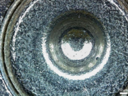
The forensic examiners not only compare projectiles, but also cartridge cases found at the crime scene. Here too, they apply the comparison principle by test firing a potential crime weapon. There are three main traces typically found on a cartridge case. One of them is the firing pin impression formed when a pin hits the primer and makes the cartridge explode. The experts also examine the breech mark – the impression made when the cartridge is pressed against the end of the barrel during the explosion – and the ejector and extractor mark caused when the cartridge is discharged or ejected from the barrel.
.")

Burglars’ tools leave traces, too
Another main application of comparison macroscopes is the forensic examination of so-called tool traces. Frequently, a perpetrator uses a tool to break or prize something open – such as a screwdriver to force a door open. This always leaves highly characteristic marks on the object. Even when brand-new, a screwdriver has a totally characteristic tip. The more often it is used, the more unique it becomes: edges chip off, it acquires scratches and grooves. Using a special silicone paste, the experts make a negative imprint of the damaged door, for example. Here too, the underlying principle is comparison, that is to say, they need a tool that could have been used for the crime. They press the screwdriver found in the suspect’s home in lead plates at various angles. As lead is a soft metal, the characteristic of the tip is obtained in a long drawn-out mark. Then they make another negative imprint of this lead plate before comparing the two silicone imprints – from the crime scene and the lead plate – under the macroscope.
Faked documents under the stereomicroscope
Documents, another area of forensics, are also inspected through a macroscope or stereomicroscope – though not necessarily a comparison instrument. The experts examine the authenticity of documents of all types, such as checks, wills or ID cards. An important part of document inspection is ink analysis. In the case of a sheet of paper printed in a laser printer, the whole sheet, even the part that looks white to the human eye, is sprayed with microscopically small particles from the toner. These are spread over the entire sheet. If someone manipulates the sheet of paper, i.e. writes something over the original, he presses these toner particles down with a biro, say. This tells the forensic expert that the document has been altered later. The comparison microscope is used, among other things, for looking at adhesive strips from a letter bomb parcel, and comparing them with a roll of adhesive tape found at a potential perpetrator’s.
.")

Microscopy brings invisible clues to light
So macroscopy and stereomicroscopy are suitable for relatively large samples like ammunition parts, toolmarks and document analysis. Here it is sufficient to examine the weapon or tool under a macroscope to be a hundred per cent certain whether or not it was used for a particular crime. Traces that are invisible to the human eye, like hairs or fibers, are investigated under a microscope at magnifications of up to 1000x, although microscopy is often only one step of several in the process chain for such small samples. The comparison microscope has a comparison bridge that rests on two full-fledged microscopes. This distinguishes it from the comparison macroscope, which has two sets of optics in one instrument.
Crimes always leave fiber traces behind, too. For instance, they can be detected on a chair four weeks after a person sat on it. The crime scene techs use large adhesive foils to remove the fibers. First they have to decide which fibers are relevant. Here again, this depends on finding an article of the suspect’s clothing that could match the fibers at the crime scene. Initial classification with a magnifier or stereomicroscope is followed by examination of the two fibers under the comparison microscope. Even brand-new clothes are never identical, because they are immediately exposed to different environments. They soon develop their own particular signature due to the fact that the fibers have faded in sunlight, for instance. These differences or similarities can be detected under the comparison microscope. However, the capacity of the human eye to notice color differences in fibers is limited. Finally, the specialists use photometric techniques to conclusively prove whether the fibers from the crime scene and those from the suspect’s clothing really are identical.

Hairs: DNA analysis delivers ultimate proof
The procedure is similar for hairs found at the crime scene. They are first classified with a magnifier and stereomicroscope and then examined under the comparison microscope. Again, the forensic experts compare hairs from the crime scene with those of a potential suspect. First they view them through the microscope to make sure they are human hairs. If so, they determine the hair type. Is it Asian, Caucasian or African? This and many other questions can be answered by studying the so-called medulla – a thin black line that runs through every hair like a spine. As with fiber examination, microscopy is often not the last step in the examination process. Another method for clearly identifying hairs is DNA analysis.

Paint traces left after hit-and-run crashes: Internationally valid color charts make work easier
Another large area of forensics is the examination of traces from cars such as glass and artificial glass splinters, paint traces and turn signal parts that gain relevance after hit-and-run accidents. The criminal investigation offices have color charts of all car paints available worldwide that enable them to allocate any paint to a specific vehicle type and the year it was made. When car manufacturers introduce a new color for a new model, they always have to immediately provide the criminal investigation offices worldwide with a color chart. If the forensic team at the scene of an accident find paint traces, for instance, that can be assigned to a specific type of vehicle, they start to investigate further: Has anyone seen a damaged vehicle of this color and age? Has it been repaired in a garage or paintshop? Here again, if the detectives find a potential crime vehicle, they compare the two paint samples under the comparison microscope. However, such investigations are only conducted if the case is fairly serious – as with car accidents involving injured persons, for example.
So forensics comprises many different areas that have a lot in common. Basically, it is always a matter of comparison – both macroscopic and microscopic. And all forensic investigations stand or fall on obtaining a hundred per cent certainty: Whether examining ammunition parts, fiber traces or paint traces, the experts have to prove without a doubt that the two samples either match or definitely do not match. Otherwise, as a piece of evidence, the clue is worthless.


")
