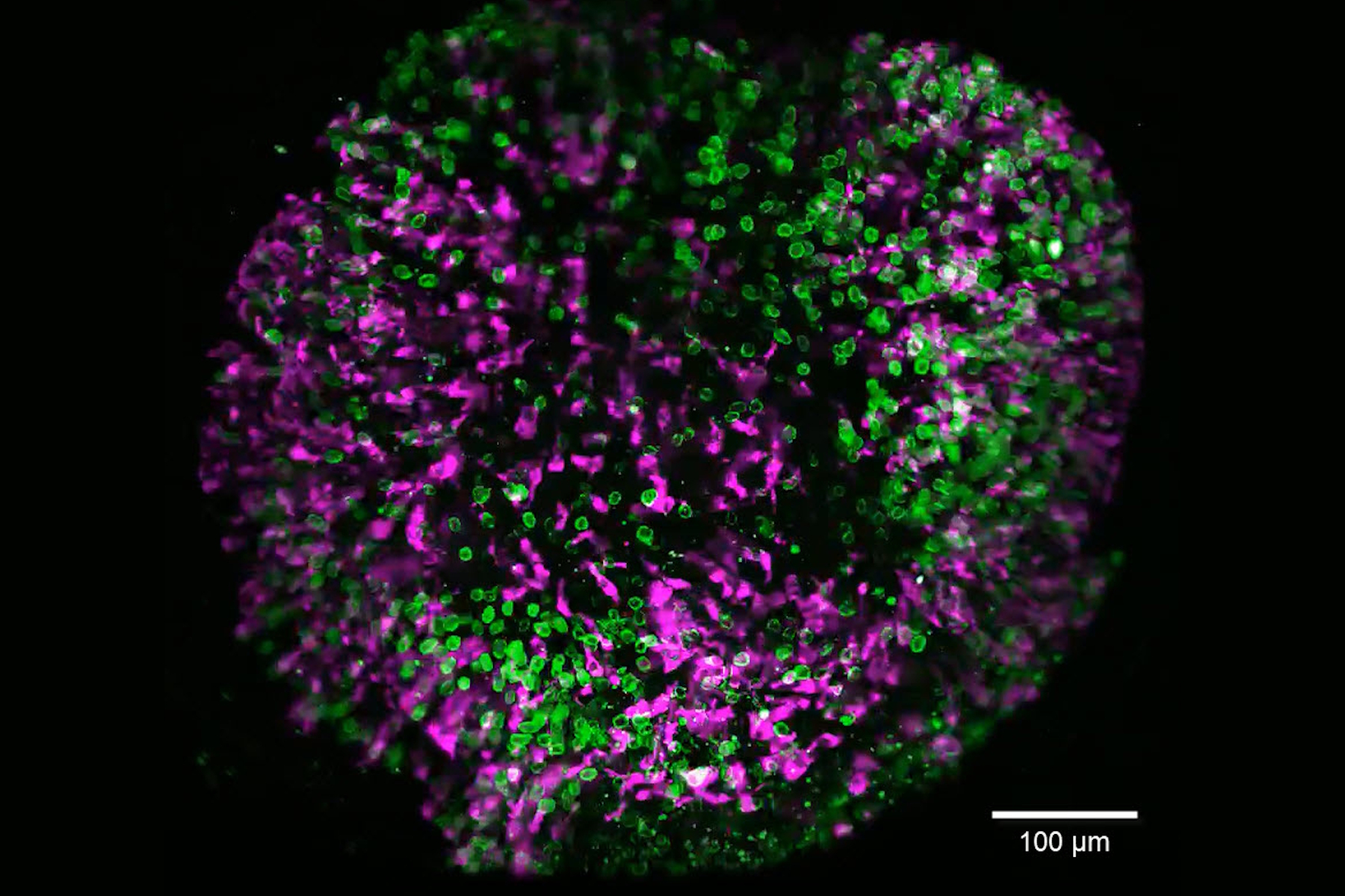
 and tubulin (magenta), acquired using Viventis Deep. Courtesy of Akanksha Jain, Treutlein Lab ETH-DBSSE Basel (Switzerland).")
The evolving landscape of drug discovery
3D culture systems such as organoids are seeing increasing use, driven by scientific innovation and regulatory shifts such as the FDA’s Modernization Act 2.0 [1], which encourages the adoption of new approach methodologies (NAMs) and non-animal technologies (NATs).
The physiological relevance of organoids allows them to mimic human tissue architecture, offering insights into dynamic cellular interactions and spatial organization. By enabling more predictive, ethical, and cost-effective research, organoids are helping to improve translational success in biomedical research, which will ultimately accelerate drug development.
Overcoming barriers in organoid and spheroid imaging
Long-term, live-cell, imaging of dense 3D samples such as organoids and spheroids presents several unique challenges compared to imaging of traditional 2D models. To fully harness the potential of these physiologically relevant 3D models, specialized imaging approaches are needed to capture and quantify complex processes in real time.
In this eBook, we outline some of the challenges encountered when imaging 3D models and explore how these can be overcome with optimized workflows including advanced imaging technologies and AI based-analysis, including autonomous microscopy.
Real-life case studies
The examples in this eBook highlight real-world applications of how our imaging solutions are helping
to advance discovery with organoid imaging. They include:
- Imaging lung organoids at the “liquid-air interface” to study potential therapeutic approaches for lung disease
- Investigating the emergence and maturation of secretory cells in intestinal organoids for spatiotemporal characterization of organoid development
- Advancing uterine regeneration therapies with endometrial organoids
- Imaging anti-cancer drug uptake in spheroids

, tubulin with Cy5 (red), and nuclei with DAPI (blue). Image courtesy of Dr. Günter Giese, Max Planck Institute for Medical Research, Heidelberg, Germany.")