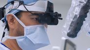
![High-resolution 3D optics [JB19.1]with enhanced depth of field [JB20.1]allow surgeons to clearly identify and safely dissect perforators and perform delicate vessel anastomoses. Mr Dariush Nikkhah. DIEP surgery London.](/fileadmin/_processed_/8/c/csm_Bilateral_DIEP_flap_breast_reconstruction_Ghali_and_Nikkhah_788764c700.jpg)
![High-resolution 3D optics [JB19.1]with enhanced depth of field [JB20.1]allow surgeons to clearly identify and safely dissect perforators and perform delicate vessel anastomoses. Mr Dariush Nikkhah. DIEP surgery London. Bilateral_DIEP_flap_breast_reconstruction_Ghali_and_Nikkhah.jpg](/fileadmin/_processed_/8/c/csm_Bilateral_DIEP_flap_breast_reconstruction_Ghali_and_Nikkhah_eda5f2f8ca.jpg "Advanced microsurgical visualization in practice: bilateral DIEP flap breast reconstruction performed by Prof. Shadi Ghali and Mr Dariush Nikkhah.")
Introduction
DIEP flap breast reconstruction is an autologous microsurgical procedure that depends on precise vessel dissection, anastomosis and perfusion assessment [1]. This article discusses how surgical microscopy and ICG with fluorescence imaging can support visualization, perfusion evaluation, workflow and team coordination during complex reconstructive microsurgery.
What is DIEP flap breast reconstruction?
DIEP (Deep Inferior Epigastric Perforator) flap reconstruction is a form of tissue transplantation that uses skin and fat from the lower abdomen (similar to a tummy tuck) to reconstruct the breast after mastectomy. Crucially, it preserves the abdominal muscles, instead dissecting out tiny perforator vessels (deep inferior epigastric vessels) and reconnecting them to the internal mammary vessels in the chest using microsurgery [1].
This leads to more natural results compared with implant-based reconstruction and avoids problems such as capsular contracture, implant-related complications, malposition, rupture, and chronic pain [1,2,4]. The DIEP flap also has the added advantage of lower donor-site morbidity, and faster functional recovery compared to older muscle-sacrificing techniques like TRAM (Transverse Rectus Abdominis Myocutaneous) flaps. And now with the advent of minimal access DIEP flap and robotic harvested DIEP flaps, patients can be discharged discharge (48 to 72 hours) and expect faster recovery [3,6,7]. Our group, in a series of 33 flaps (15 MFI and 18 standard incisions) identified advantages to minimal access DIEP [3]. The median length of stay was significantly lower in the MFI group [4 days (IQR 4-5)] vs. [5 days (IQR 4-6.25)], (p=0.036). Mesh use was significantly higher in the standard incision group (p=0.001). There was no significant difference in operative time (p=0.128). The average MFI rectus sheath incision length was 4.59 cm. No donor-site complications occurred in either group [3].
Robotic DIEP surgery is still in its infancy. Recent systematic reviews have demonstrated that it may reduce postoperative pain and limiting abdominal donor site morbidity [5,6]. Potential limitations include longer operative times, variable hospital stays, and increased costs [5,6]. Our group has adopted the transabdominal preperitoneal (TAPP) approach, and we have demonstrated that short fascial incisions can be achieved with the advantages of a long pedicle and easier access to the deep inferior epigastric vein and artery as opposed to the hand assisted technique [6,7]. The mean robotic operative time in this review between centers was 64 minutes, with total operating times averaging 574 minutes for TAPP and 497 minutes for TEP. The mean length of stay was 5 days, and the mean fascial incision length was 3 cm [6]. This is comparable to current non-robotic techniques in DIEP flap reconstruction [6].

The trade-off of microvascular breast reconstruction is that it is technically demanding, requiring precise dissection of small vessels and meticulous microvascular anastomosis [1]. These cases are often performed by two plastic and reconstructive surgeons to minimize operative time with one surgeon preparing the recipient vessels in the chest and the other surgeon raising the flap.
What are the advantages of using a surgical microscope?
Some groups utilize high magnification loupes to perform microvascular surgery. However, we find that this approach has several disadvantages and does not allow for the high level of precision during microsurgery which can lead to errors resulting in vascular thrombosis and potential transplant failure.
How is the ARveo 8x surgical microscope valuable in the context of DIEP flap microsurgery?
Microvascular visualization:
High-resolution 3D visualization with enhanced depth perception can support surgeons in identifying perforators and performing microsurgical procedures such as vessel anastomoses [1]. (See Video #1). The venous anastomosis of the DIEP flap is performed using a Synovis coupler end to end onto the internal mammary vein, this enables rapid anastomosis of multiple superficial and deep veins preventing flap congestion.
Video 1: High-resolution 3D optics with enhanced depth of field allow surgeons to clearly identify and safely dissect perforators and perform delicate vessel anastomoses. Mr Dariush Nikkhah. DIEP surgery London.
The arterial vessel anastomosis of the DIEP flap is performed by hand using a posterior wall technique end to end onto the antegrade internal mammary vessel; 8.0 nylon sutures are used to bring the vessel ends together. The posterior wall technique is useful in deep spaces such as the chest and is our standard approach in free tissue transfer.
Intraoperative perfusion assessment:
Fluorescence imaging [e.g., ICG] enables intraoperative visualization of flap perfusion and vessel patency without interrupting the surgical workflow (See Video #1) [8]. We found that this enables us to visualize perfusion through the arterial anastomosis and provides information that supports assessment of vascular flow. Intraoperative Indocyanine green (ICG) angiography can provide real-time information on tissue perfusion during breast reconstruction [9]. In DIEP flap reconstruction, a 2023 systematic review by Wang and co-authors reported fat-necrosis rates of 10.9% with ICG angiography compared with 21.5% using conventional assessment [9].
Video 2: With the GLOW800 AR fluorescence application and ICG, surgeons can visualize vessel patency and tissue perfusion in real time, complemented by 3D visualization to support breast reconstruction procedures.
Performance in deep operative planes:
The advanced illumination, high-resolution visualization, and depth perception of the ARveo 8x surgical microscope support visualization during perforator dissection and deep chest dissection when preparing the internal mammary vessels. (See Video#4). Based on my experience, the super microsurgery mode has been particularly valuable when performing delicate procedures such as perforator-to-perforator anastomosis in free tissue transplantation [10].
Video 3: Perforator dissection is one of the most delicate stages of DIEP flap breast reconstruction. Watch Mr Dariush Nikkhah demonstrating the importance of advanced surgical visualization for identifying and preserving critical vascular structures within delicate tissue planes.
Ergonomics for long, complex cases:
The microscope has a small base and can fit in the operating room with other devices such as robotic systems as Da Vinci Robot used for DIEP flap harvest [5,6]. Furthermore, the heads-up display options and flexible viewing of ARveo 8x support ergonomic working positions during lengthy DIEP procedures, helping surgeons remain comfortable throughout the procedure.
Video 4: DIEP flap breast reconstruction, procedures commonly last 6–8 hours, and bilateral reconstructions can take even longer. The ARveo 8x 3D surgical microscope frees surgeons from traditional microscope eyepieces with integrated 3D visualization displayed on a 55-inch heads-up monitor, supporting an ergonomic workflow during long surgery.
Improved team coordination and teaching:
The large display on the microscope and the shared 3D visualization allow assistants to follow critical microsurgical steps in real time. This can be used in live surgery demonstrations and teaching resident doctors.
We have also noted that the primary scrub nurse can better follow the relevant microsurgical steps in this procedure.
Video 5: In this video, Dr Dariush Nikkhah shares real-time fluorescence imaging of blood flow with the team, highlighting the progressive filling of veins following microvascular breast reconstruction. The shared 3D visualization provided by ARveo 8x allows the entire surgical team to follow critical microsurgical steps.
Conclusion
DIEP flap reconstruction demands a high level of precision and careful intraoperative visualization. In my experience, ARveo 8x supports these procedures through high-quality magnification and visualization capabilities, while ICG imaging with GLOW800 enables real-time visualization of blood flow during microsurgical breast reconstruction. These visualization tools can support intraoperative assessment and decision-making, particularly in complex cases such as patients who have undergone radiotherapy. In my clinical experience, the use of enhanced magnification together with ICG and GLOW800-assisted visualization has supported intraoperative assessment of blood flow and perfusion during microsurgical breast reconstruction [2].