
Continuous advances in microsurgery enable complex breast, and head and neck reconstructions for cancer patients. These reconstructions largely rely on free flap techniques and aim for both functional and aesthetic rehabilitation of the patient [1,2]. Most used free flap procedures include [3]:
| Head and neck reconstruction | Breast reconstruction |
|---|---|
| Fibula osteocutaneous flap | Deep inferior epigastric perforator (DIEP) flap |
| Radial forearm free flap (RFFF) | Latissimus dorsi (LD) flap |
| Anterolateral tight (ELT) flap | Rectus abdominis (RA) flap, vertical (VRAM) or transverse (TRAM) |
Challenges during reconstructive surgery
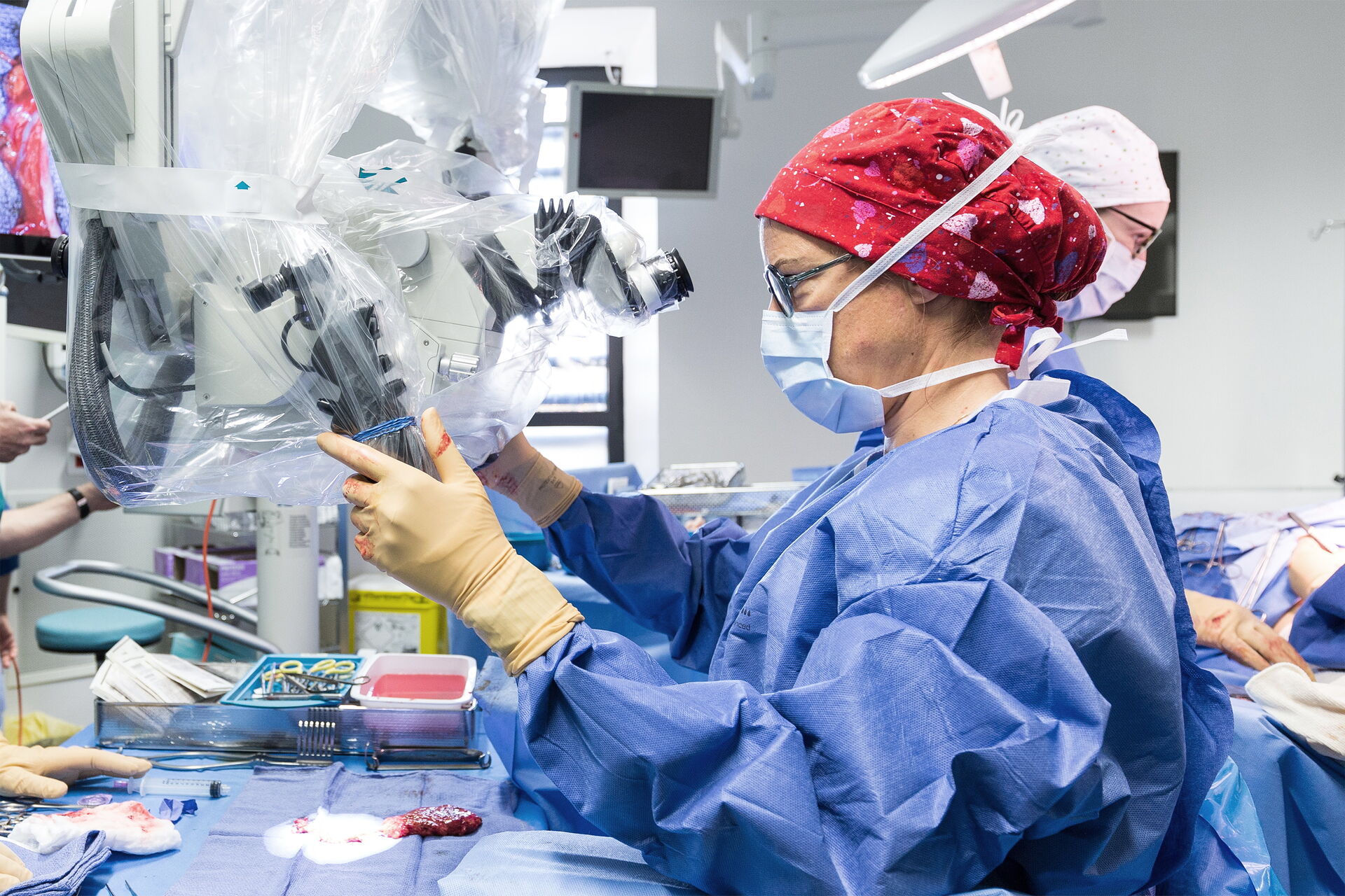
- Given the long operating time needed for these procedures, plastic surgeons are at a higher risk of developing musculoskeletal problems, due to awkward positioning, hyperflexion of the cervical spine and repetitive motions [4].
- Moreover, meticulous microsurgical technique is essential for successful flap reconstructions and a clear, sharp view of the operating field is key to facilitate the reconstruction procedure.
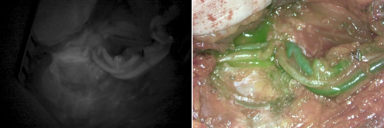
- Flap necrosis is one of the most common complications following reconstruction. The use of fluorescent angiography to assess blood flow and tissue perfusion reduces the rate of flap necrosis and flap loss, contributing to the success of the reconstruction [5].
- Microsurgical microscopes with integrated fluorescence cameras allow all-in-one visualization, streamlining the surgical workflow without interruptions. ln addition, new Augmented Reality technology provides
unaltered visualization of anatomy and physiology without the need to divert the eyes from the operating fieId [6].
Enhanced ergonomics
- Easily adjust working distance, zoom and field of view, for both close visualization of anastomosis and wide view of flap perfusion, in high resolution, without letting down your surgical tools.
- Find the ideal operating position - through the eye pieces or heads-up surgery - and limit musculoskeletal issues for you and your assistant team. Follow every step of the surgery on HD/4K screens, in 2D or 3D.
- Ensure an optimized workflow with microscopes specifically designed for long arm reach and small footprint inside the OR.
Outstanding optical features
- See more and progress confidently with FusionOptics™ - unique combination of high resolution & enhanced depth of field.

Figure 1: FusionOptics TM technology.
- Boost magnification by 40% with the Magnification Multiplier.
- Capitalize on our automatic optimization of light intensity - BrightCare - to minimize incidents and reduce tissue damage.
Groundbreaking visualization technology
- Easily check the flap viability with indocyanine green (ICG) thanks to the integrated FL800 fluorescence option.
- Push the boundaries of microsurgery with our patented built-in GLOW800 Augmented Reality (AR) - further enhance the natural color visualization of anatomy augmented by real-time view of vascular flow and tissue
perfusion, to guide your surgical decisions with confidence.