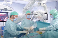

Continuous real-time view of the surgical field
Neuronavigation works like a GPS system, enabling accurate position tracking of instruments within the patient’s anatomy. Surgeons can outline the tumor, identify organs at risk, and define the optimal access pathway to the tumor to prevent injury to healthy structures.
Before surgery, CT or MR images of the patient are taken. In the operating room, the navigation system matches these images up with the live patient images using a wireless laser pointer. The pointer identifies surface points on the patient’s skin which are transmitted to the navigation system via an infrared camera. During the procedure, the camera also transmits the locations of the instruments and equipment – including the microscope – which are outfitted with reflective markers.
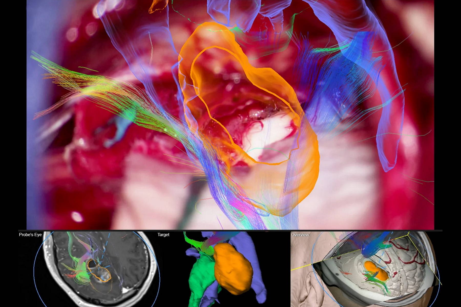
The patient images prepared prior to surgery are loaded into the navigation system and reconstructed into images on the monitor or injected into the oculars of the surgical microscope. This gives the surgeon a continuous, real-time map of the position of the instruments in relation to the brain structures and the tumor.
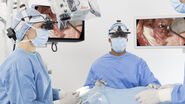
Microscope integration offers new imaging options
Brainlab is one of the market leaders in the field of image-guided technology and was the first to work together with Leica to integrate its neuronavigation software with surgical microscopes. With improved 3D visualization, the new Brainlab software provides a more realistic representation of planned objects, structure and fiber tracts throughout the entire procedure.
Touch-based rotation of the microscope view on the navigation screen allows for a look behind the actual video plane to reveal underlying structures in 3D for more anatomical insight. The “Probe’s Eye” feature reconstructs the patient dataset to the microscope’s position and orientation for a direct comparison of preoperative data and intraoperative anatomy. With “Navigation update” the surgeon is able to correct initial registration based on the patient’s anatomical landmarks.

Limiting guesswork in tumor resection
Every neurosurgical procedure demands a vast amount of surgeon experience, patience and competence. Although neuronavigation does not replace surgical skill, it effectively supports the surgeon in pre-operative planning and during the procedure by giving more precise information in the most critical moments.
"The integration of the microscope with neuronavigation helps the surgeon to accurately identify questionable areas", explains Valentin Elefteriu, Product Manager Augmented Reality at Brainlab. "For example, if they focus on the edge of a tumor and can’t tell exactly where the tumor tissue ends, a look at the pre-planned anatomic data with microscope image overlay reveals whether the focused area is part of the tumor or not. Precise planning may also minimize head shaving, skin incisions, and bone flap sawing and thus reduce stress for the patient.”
Brainlab is a registered trademark of Brainlab AG registered in Europe, the United States and other countries.
 case.")