
The dental surgical microscope has become increasingly important for high-quality and successful dental medicine, particularly in the field of endodontics. A dentist can conduct micro-invasive surgeries which aim to preserve tooth substance, conserve tissue, minimize risks, and reduce healing time with the help of a microscope. To choose the microscope that best fits the dentist’s needs, it is helpful to know some of the decisive features of a modern dental microscope described in this article.
Optical quality
A dental microscope is at its core an optical instrument, which allows the dentist to see enhanced detail in order to work as precisely as possible. Therefore, there should be no compromise concerning the optical components. Only superior optical quality with excellent clarity offers high resolution, large depth of field, and maximum light transmission.
For successful root-canal treatments, a dental microscope is indispensable as root canals are, in most cases, not straightforward. There are often cavities and tiny ramifications which are difficult to detect without a high magnification and large depth of field. Accurate color representation is also required for easy differentiation of anatomical details. Optics with apochromatic correction, standard with the M320 dental microscope, provide images without chromatic (color) aberration.
Video: Dental videos recorded by the M320 dental microscope with integrated 4K camera from Leica Microsystems on endodontic surgery. Video courtesy of Dr. Alessandro Patisso from Francavilla, Fontana BR, Italy.
Illumination
In combination with optical quality, illumination plays a key role in terms of brightness and color of the image that the dentist sees through the eyepieces or on an electronic monitor. For natural color representation, light with a color temperature between 5,000 and 6,000 kelvin (K) is optimal. The M320’s efficient and powerful LED illumination ensures high color perception and reproduction enabling easy differentiation of anatomical details.
Another factor to consider is the lifespan of the light source as this impacts the cost of ownership, potential downtime, and environment. Compared to xenon or halogen light sources, LED illumination usually lasts much longer. A longer lifetime reduces the frequency of light-source exchange and, therefore, potential microscope downtime.
The location of the light source may also influence its life span. When the light source is located in the microscope stand, the light needs to be transmitted with a fiber optic cable through the arm to the optics carrier.
The fiber optics can be subjected to wear and tear through everyday microscope adjustments which, over time, could impact the light reaching the treatment site. The M320 is designed to have the LED illumination directly in the optics carrier to avoid this potential cause of brightness degradation.
Documentation
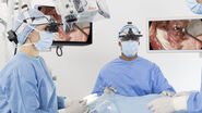
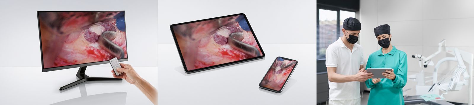
Today, video and images play an ever-increasing role within the dental practice for a number of reasons. Firstly, during a procedure, live on-screen video can help the assistant to better support the dental surgeon. Video is also a useful training tool either during or post treatment and can be additionally shared with the wider dental community online, such as with social media, or at seminars.
Additionally, video can support patient relationships and trust as the dentist shows the patient what they see, takes them through the steps of a procedure and, thus, includes them in consultation and treatment.
Finally, video and images can be included in a patient’s file for more thorough documentation and to facilitate easy review. Ease of transfer to a practice documentation system and the ability to save different file formats are, therefore, further key considerations.
The M320 dental microscope has an integrated UHD 4K camera and only requires a push of a button for still or video capture, with no interruption of the workflow. Data can be saved to a SD memory card or transferred directly via USB.
Ergonomic design
Musculoskeletal pain caused by a hunched working position and harsh, repetitive movements, can have a serious impact on the dentist’s professional and personal life. Using a microscope during dental procedures helps the dentist to maintain a neutral, upright working position. Modern microscopes should also offer a variety of ergonomic accessories to adapt the microscope to the user’s individual body frame and working preference.
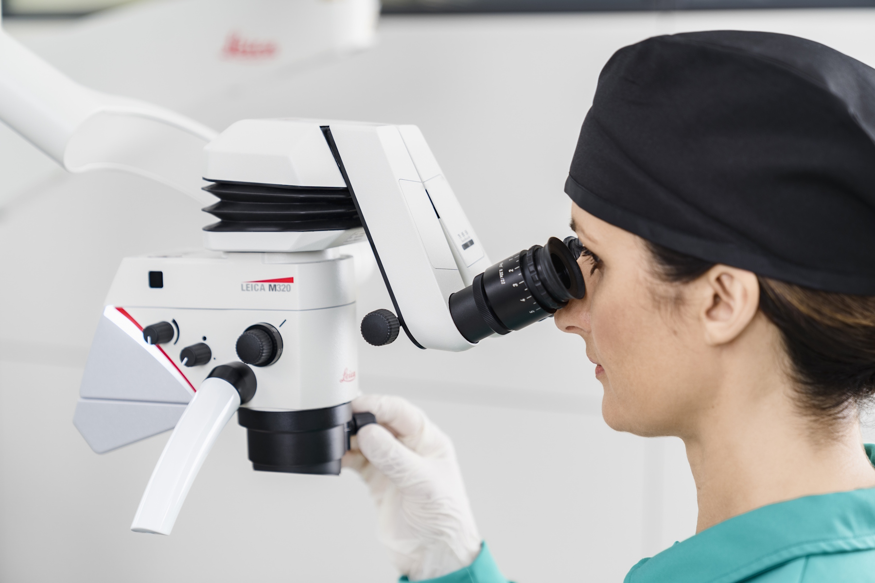
The M320 dental microscope features two binocular tubes with a 45˚ angle for simple procedures and another with 180˚ movement for increased positioning flexibility. The ErgoWedge accessory supports positioning ease and the ErgonOptic Dent accessory further extends the reach of the microscope and provides a greater range of movement by allowing the optics carrier to swivel to the angle needed while maintaining an ergonomic posture.

Workflow integration
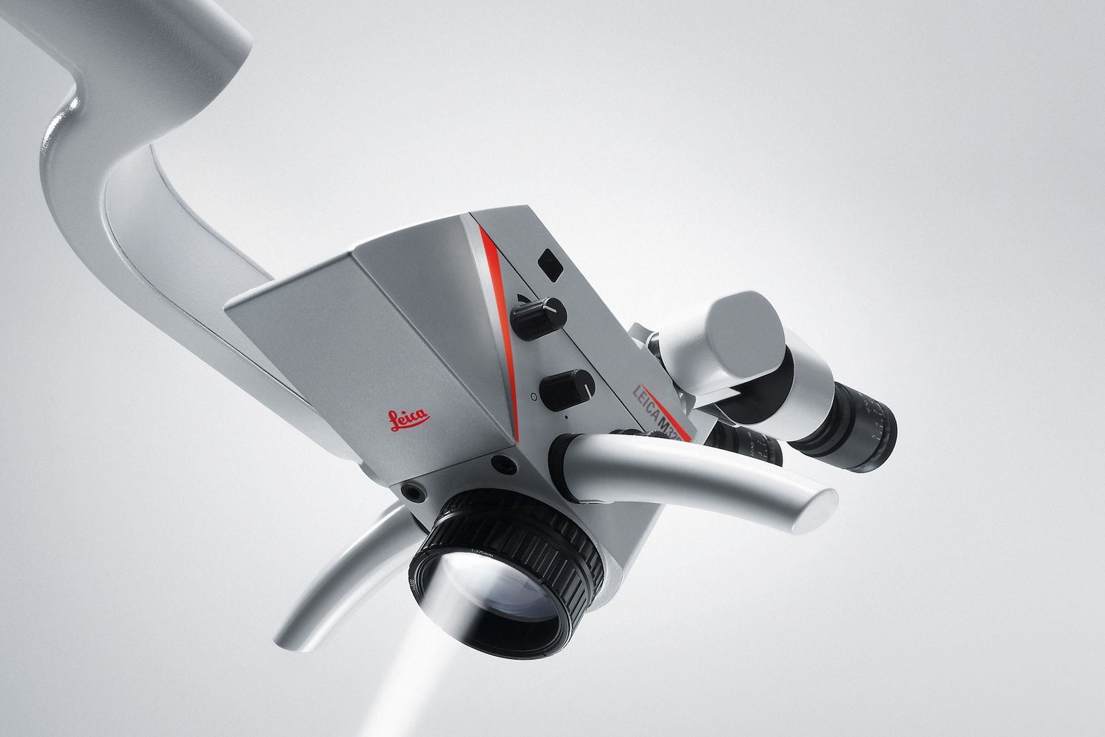
The design of a microscope takes into account both aesthetics and functionality. A carefully considered design should make it easy to integrate the microscope into the practice environment and even improve the workflow. The M320 was designed with the individual needs of dentists in mind: its streamlined design with various mounting options, including the possibility to integrate with several treatment units, allows it to be tailored to the individual demands of each practice.
Quick, lightweight, and accurate positioning combined with fast stabilization supports a smooth workflow. The optional MultiFoc Objective with a variable working distance from 200 mm to 300 mm allows the dentist to refocus quickly and easily as necessary. Being able to change between different focus levels during examination and treatment without having to reposition minimizes workflow interruptions. This capability can help the dentist to maintain efficiency and concentration.
Hygiene
In a dental practice, a high standard of cleanliness is a prerequisite, and every piece of equipment should make it as easy as possible to achieve this requirement. A streamlined design with cables routed internally from the optics carrier to the stand, facilitates cleaning and also avoids accidental cable damage. Additionally, M320 dental microscopes are coated with a paint, which is designed to provide an antimicrobial effect on surfaces.