
Microscope Solutions for Cornea Surgery
Corneal diseases are the third leading cause of blindness wordwide1. They can be congenital or caused by injury, trauma, infection or allergy, resulting in loss of transparency and opacities. According to the World Health Organization, 4.2 million people globally are affected by corneal opacities2.
Some corneal opacities can be treated with eye drops and medication. However, when the opacities are too thick and the cornea severely damaged, corneal surgery may be necessary to restore vision. A corneal transplant is performed to replace the damaged part of the cornea with healthy corneal tissue from a donor. Each year, approximately 185,000 corneal transplants are performed in 116 countries3.
Contact a local specialist for expert advice on the right solution for your needs and budget.
Cornea Surgery Procedures
There are three main corneal transplant surgical methods: penetrating keratoplasty (PK); endothelial keratoplasty (EK) including Descemet stripping automated endothelial keratoplasty (DSAEK) and Descemet membrane endothelial keratoplasty (DMEK); and anterior lamellar keratoplasty (ALK) which can be superficial (SALK) or deep (DALK).
Corneal transplants are highly specialized procedures, requiring advanced surgical skills and experience to avoid complications and graft failure. The use of a cornea microscope helps surgeons achieve optimal visualization to operate in the best conditions.
Cornea Surgery Challenges
Cornea surgery has evolved considerably in recent years, with corneal lamellar procedures largely replacing penetrating keratoplasty. Tissue grafts also tend to be thinner and thinner to achieve better visual acuity and lower the risk of rejection4.
Some of the biggest challenges for corneal surgeons include:
- Visualizing clearly the full anterior chamber, Descemet membrane peeling and endothelium
- Measuring how deep to cut the corneal stroma and quantifying the incision depth
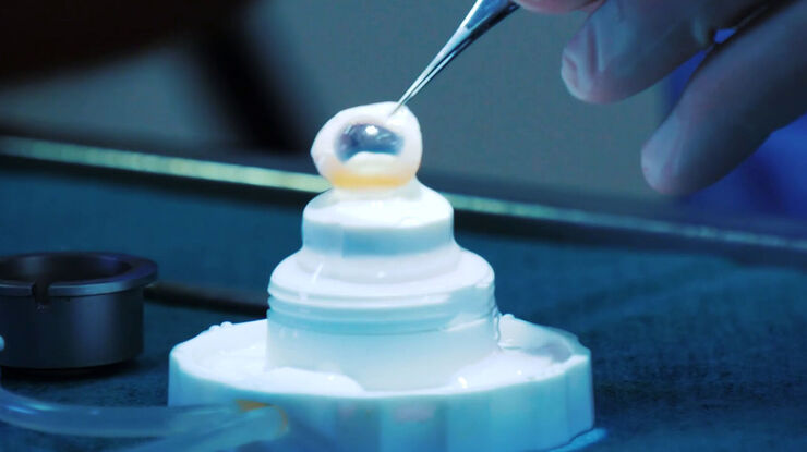
- Avoiding damage to the donor tissue
- Achieving correct unfolding of the graft within the anterior chamber
- Ensuring correct orientation and graft positioning
- Confirming complete graft attachment without folds or liquid
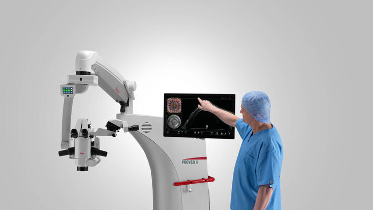
Key Features of Leica Microscope Solutions for Cornea Surgery
Powerful illumination
Benefit from homogenous light distribution and optimal light transmission for graft preparation and transplant management.
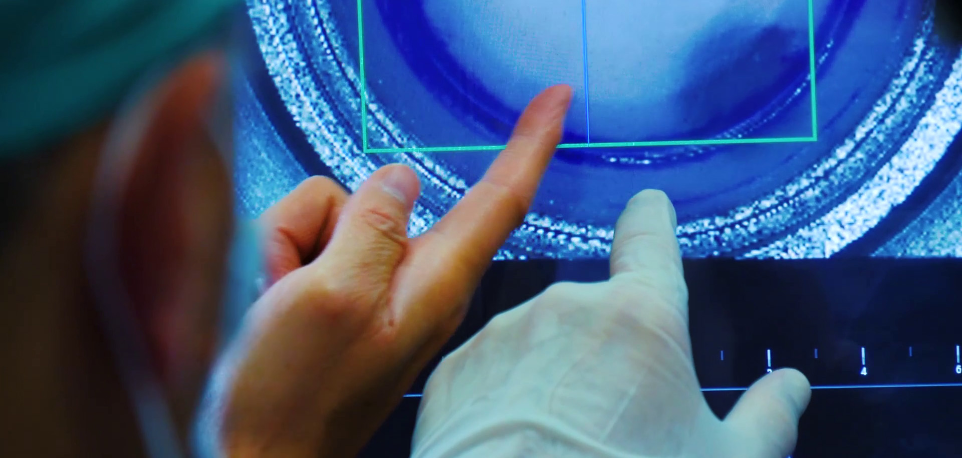
Real-time insights
Obtain real-time intraoperative confirmation of how tissue reacts to surgical maneuvers to achieve optimal graft orientation and positioning.
Exact measurements
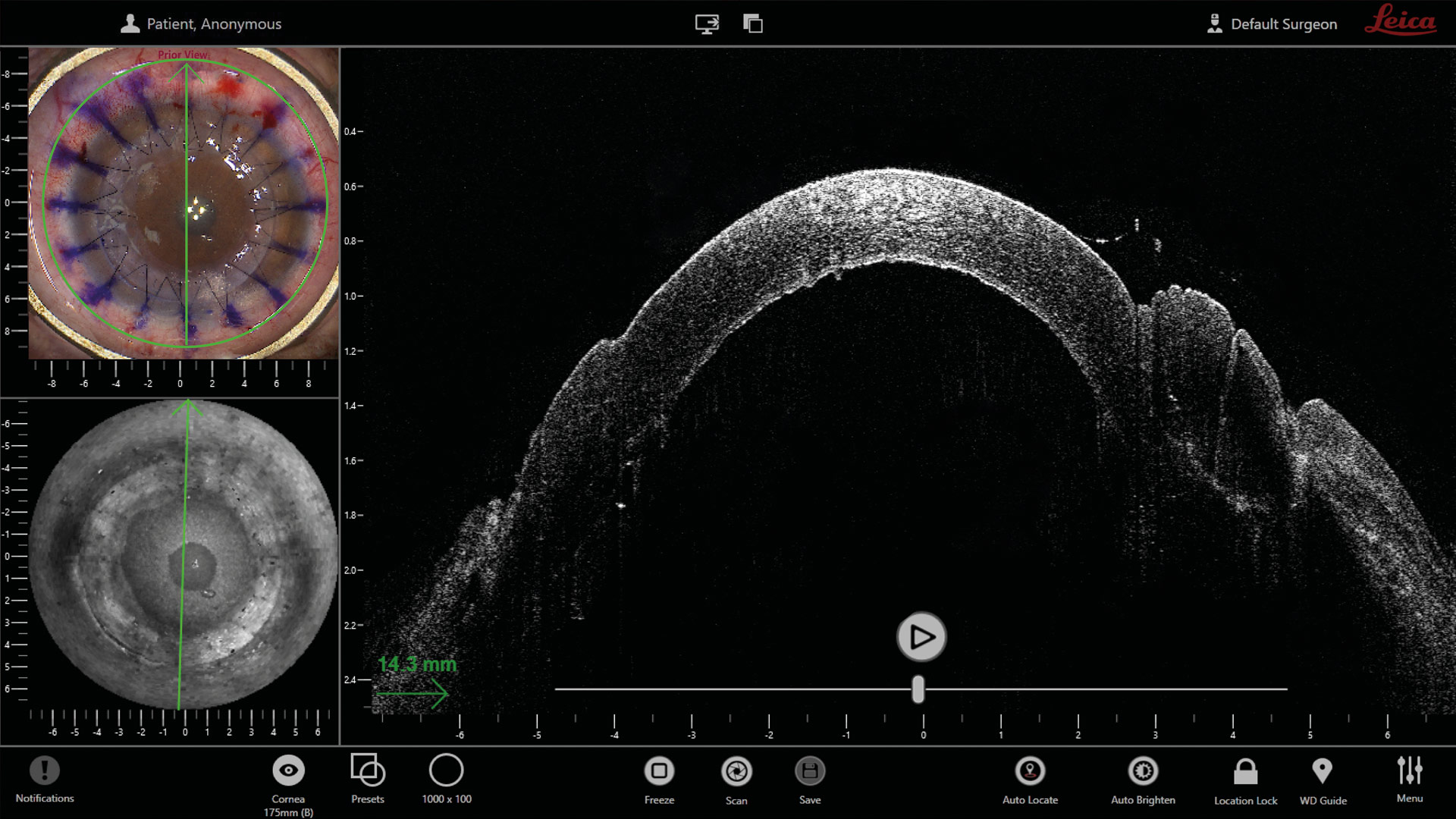
Leverage intraoperative OCT for measurements of corneal thickness, depth of the inserted cannula during air injection and trephination depth.
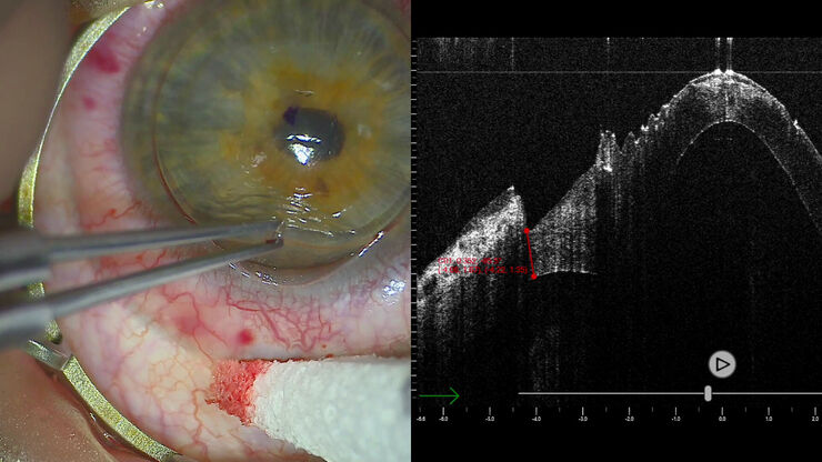
In this video, see how high-resolution intraoperative OCT can enhance visualization during DMEK surgery with real-time, cross-sectional imaging of subsurface details.
Cornea Microscope Solutions: DMEK surgery with EnFocus intraoperative OCT
During DMEK surgery, accurate donor membrane positioning and full adherence to the stroma are crucial to patient outcomes. EnFocus intraoperative Optical Coherence Tomography can reveal more information to support each stage of the procedure.
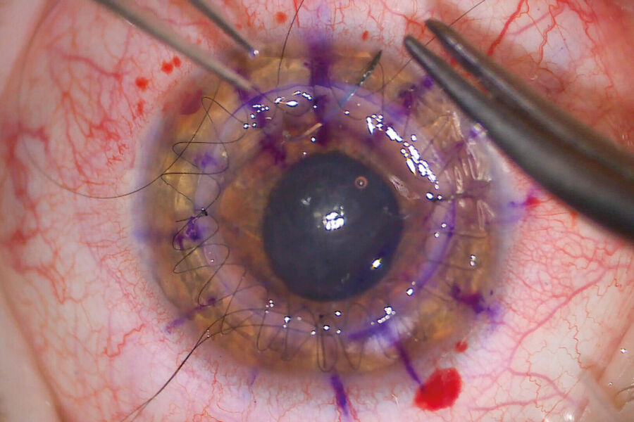
Corneal Perforation and Trauma Repair Procedures
Corneal perforations can be challenging to manage, even for seasoned corneal surgeons. Intraoperative Optical Coherence Tomography can provide useful insights for ocular perforation and trauma procedures. It can help assess the anatomy and extent of the damage, and support repair.
Frequently Asked Questions Cornea Surgery
A cornea transplant is an operation to replace damaged corneal tissue with healthy corneal tissue from a donor.
Cornea transplant is the current standard treatment for corneal blindness.
The majority of cornea surgery operations are successful. However, rejection occurs in about 10% of cornea transplants5. There may also be complications during surgery.
Many corneal conditions can be treated effectively with corrective lenses, eye drops or medication. However, if damage is too important, a corneal transplant may be required.
After obtention of the corneal tissue from the donor, the cornea is placed in a storage medium to keep the tissue viable.
Related Articles

4 Key Benefits of 3D Digital Microscopy in Ophthalmic Surgery

Expert Techniques for Superior Visualization in Cataract Surgery

Ophthalmology Case Study: Corneal Transplantation

Glaucoma Stent Revision Surgery Guided by Intraoperative OCT

A Larger 3D Area in Focus for Neurosurgical and Ophthalmic Microscopes

How Real-Time OCT Imaging Impacts Precision in Corneal Surgery?

Buying an Ophthalmic Microscope? Gain Peers Insights from Dr. Dhami

RPE65 Gene Therapy Subretinal Injection: Benefits of Intraoperative OCT

Dislocated Cataract Angle Closure Aided by Intraoperative OCT

Posterior Segment Surgery: Benefits of Utilizing Intraoperative OCT
References
-
Chaurasia SS, Lim RR, Lakshminarayanan R, Mohan RR. Nanomedicine approaches for corneal diseases. J Funct Biomater. 2015 Apr 30;6(2):277-98. doi: 10.3390/jfb6020277. PMID: 25941990; PMCID: PMC4493512.
-
World Health Organization, Word Report on Vision, October 2019. Accessed on March 27th, 2023, at: https://www.who.int/publications/i/item/9789241516570
-
American Academy of Opthalmology, Eye Wiki, Corneal Donations, February 2023. Accessed on March 9th, 2023, at: https://eyewiki.aao.org/Corneal_Donation
-
Review of Ophthalmology, Innovations in Corneal Transplants, February 2020. Accessed on March 9th, 2023, at: https://www.reviewofophthalmology.com/article/innovations-in-corneal-transplants
-
Mayo Clinic, Cornea Transplant. Accessed on March 9th, 2023, at: https://www.mayoclinic.org/tests-procedures/cornea-transplant/about/pac-20385285
