
Especialidades médicas
Especialidades médicas
Explore una completa colección de recursos científicos y clínicos adaptados a los profesionales sanitarios, que incluye opiniones de colegas, estudios de casos clínicos y simposios. Diseñada para neurocirujanos, oftalmólogos y especialistas en cirugía plástica y reparadora, otorrinolaringología y odontología. Esta colección destaca los últimos avances en microscopía quirúrgica. Descubra cómo las tecnologías quirúrgicas de vanguardia, como la fluorescencia AR, la visualización 3D y las imágenes OCT intraoperatorias, permiten tomar decisiones con confianza y precisión en cirugías complejas.

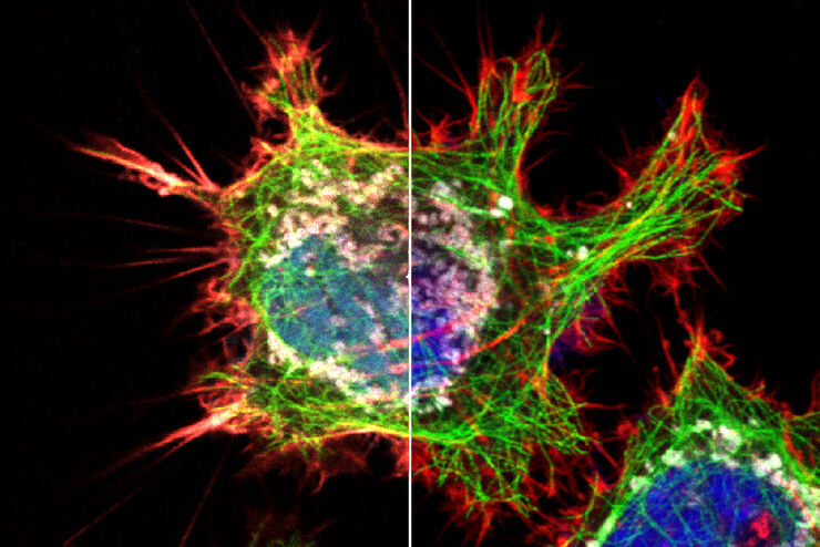
Obtenga el máximo de información de su muestra con LIGHTNING
LIGHTNING es un proceso adaptativo para la extracción de información que revela estructuras finas y detalles que de otro modo simplemente no serían visibles, de forma completamente automática . A…

Virología
¿Su interés en investigación se centra en infecciones víricas y enfermedades? Descubra cómo puede adquirir conocimientos sobre virología con soluciones de generación de imágenes y preparación de…

Microscopy in Virology
The coronavirus SARS-CoV-2, causing the Covid-19 disease effects our world in all aspects. Research to find immunization and treatment methods, in other words to fight this virus, gained highest…

Computational Clearing - Enhance 3D Specimen Imaging
This webinar is designed to clarify crucial specifications that contribute to THUNDER Imagers' transformative visualization of 3D samples and improvements within a researcher's imaging-related…
Explore Innovative Techniques to Separate Fluorophores with Overlapping Spectra
In this article we explore several strategies you can take to improve the separation of fluorophores and increase the number of fluorescent probes you can distinguish in your sample.

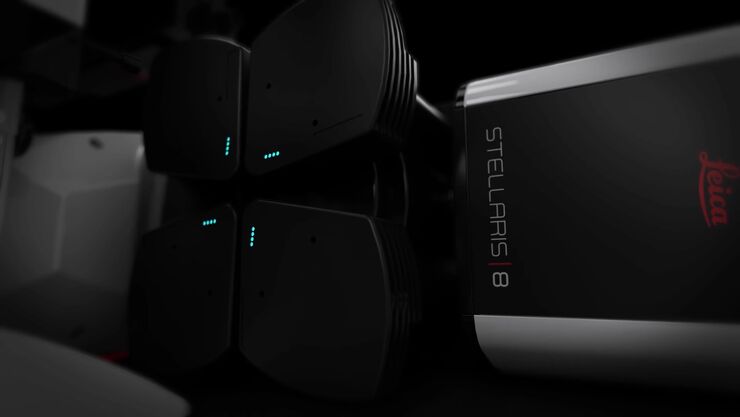
STELLARIS White Light Lasers
When it comes to choosing fluorescent probes for your multi-color experiments, you shouldn’t have to compromise. Now you can advance beyond conventional excitation sources that limit your fluorophore…

TauSense Technology Imaging Tools
Leica Microsystems’ TauSense technology is a set of imaging modes based on fluorescence lifetime. Found at the core of the STELLARIS confocal platform, it will revolutionize your imaging experiments.…
The Power HyD Detector Family
Powerful photon counting detectors on the STELLARIS confocal platform provide improved photon counting, ultra-sensitive imaging and more color options in the NIR spectrum.

Investigación del cáncer
El cáncer es una enfermedad compleja y heterogénea causada por células con una regulación deficiente del crecimiento. Los cambios genéticos y epigenéticos en una célula o en un grupo de ellas alteran…
