
Industriale
Industriale
Immergetevi in articoli dettagliati e webinar incentrati su ispezioni efficienti, flussi di lavoro ottimizzati e comfort ergonomico in contesti industriali e patologici. Gli argomenti trattati includono il controllo qualità, l'analisi dei materiali, la microscopia in patologia e molti altri. Questo è il luogo in cui potrete ottenere preziose informazioni sull'utilizzo di tecnologie all'avanguardia per migliorare la precisione e l'efficienza dei processi di produzione, nonché l'accuratezza della diagnosi e della ricerca patologica.
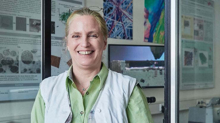
Ultramicrotome UC Enuity in Practice: Stable 15 nm Sections at ZFE
After using the UCT and UC6 ultramicrotomes, Claudia Mayrhofer calls UC Enuity a leap in stability—so robust that vibrations and temperature shifts don’t spoil sections, even with multiple users. Auto…

Eliminating Electrostatic Interference in Laser Microdissection
Electrostatic charge in laser microdissection (LMD) causes two critical failures: samples stick to charged surfaces and are lost, or samples fly into adjacent wells and cause cross-contamination. We…

History, Developments and Trends of Microscopy in Cancer Research
Cancer is a global disease, with 18 million new cases diagnosed and 10 million cancer-related deaths worldwide in 2020. This burden is set to increase, with a projected increase in cases of ~55% by…
, tubulin with Cy5 (red), and nuclei with DAPI (blue). Image courtesy of Dr. Günter Giese, Max Planck Institute for Medical Research, Heidelberg, Germany.")
Overview of Fluorescent Dyes in terms of Applications and Properties
An introduction to commonly used fluorescent dyes and an overview of their characteristics are given in this article. Fluorescence microscopy is used for the study of specific cellular components with…

Researchers Insights: Microscopy in Cancer Research
Discover how imaging techniques are driving cancer research forward. In this issue, we present comprehensive multimodal studies using microscopy, as well as new directions in intraoperative cancer…
, Tropomyosin (cardiomyocytes, red) and GFP (primordial cardiac layer, green).")
A Guide to Fluorescence Microscopy
Fluorescence microscopy uses the ability of fluorophores, dyes, or fluorescent proteins to emit light of a specific wavelength after being excited with light of a shorter wavelength. Biomolecules can…
 labeled with membrane-permeable calcein, high-pressure frozen in salt water using EM ICE.")
High-Pressure Freezing for Organoids: Cryo CLEM & FIB Lift Out
Master cryo EM workflow steps for challenging 3D samples: when to choose HPF vs. plunge freezing, reproducible blotting/ice control, contamination aware transfers, Cryo CLEM 3D targeting in organoids,…
 and astrocytes (green) in a cortical spheroid derived from human induced pluripotent stem cells.")
Guide to Live-Cell Imaging
For a wide range of applications in various research fields of life science, live-cell imaging is an indispensable tool for visualizing cells in a state as close to in vivo, i.e. living and active, as…

Factors to Consider When Selecting a Research Microscope
An optical microscope is often one of the central devices in a life-science research lab. It can be used for various applications which shed light on many scientific questions. Thereby the…
