

Development and Derisking of CRISPR Therapies for Rare Diseases
This on-demand presentation by Dr. Fyodor Urnov and Dr. Sadik Kassim, originally delivered at ASGCT 2025, focused on a critical challenge in genetic medicine: how to scale CRISPR therapies from…

Multiplexed Imaging Reveals Tumor Immune Landscape in Colon Cancer
Cancer immunotherapy benefits few due to resistance and relapse, and combinatorial therapeutic strategies that target multiple steps of the cancer-immunity cycle may improve outcomes. This study shows…
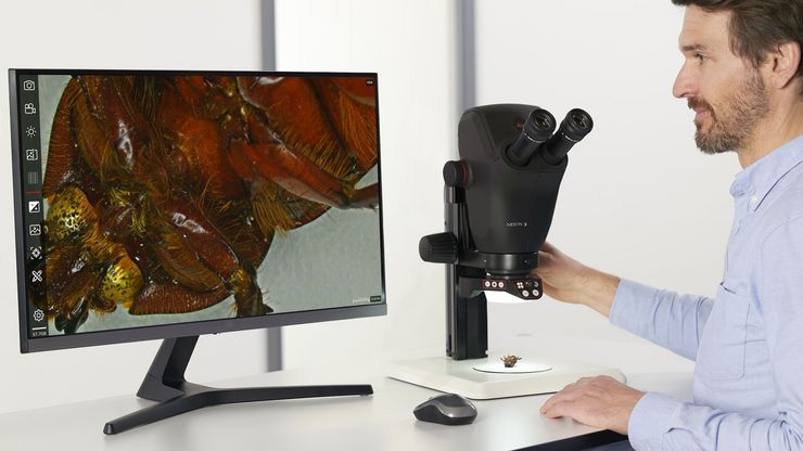
 observed with an Ivesta 3 stereo microscope during fly pushing (sorting of the flies). The scale bar length is 1 mm. Image courtesy of M. Benton, EMBL, Heidelberg, Germany.")
A Guide to Using Microscopy for Drosophila (Fruit Fly) Research
The fruit fly, typically Drosophila melanogaster, has been used as a model organism for over a century. One reason is that many disease-related genes are shared between Drosophila and humans. It is…

神経科学研究
神経変性疾患の理解向上に取り組んでいる、もしくは神経系の機能を研究をしていますか? ライカマイクロシステムズのイメージングソリューションによってブレイクスルーを起こす方法をご覧ください。

ゼブラフィッシュを用いた研究
スクリーニング、ソーティング、マニピュレーションおよびイメージングを通じて最良の結果を得るためには、細部や構造を観察して、研究の次の段階に向けて正しい判断を下す必要があります。
優れた光学系と高解像度で定評のあるライカの実体顕微鏡と透過照明スタンドは、世界中の研究者から支持されています。

Improving Zebrafish-Embryo Screening with Fast, High-Contrast Imaging
Discover from this article how screening of transgenic zebrafish embryos is boosted with high-speed, high-contrast imaging using the DM6 B microscope, ensuring accurate targeting for developmental…

Transforming Research with Spatial Proteomics Workflows
Spatial Proteomics, Nature Methods 2024 Method of the Year, is driving research advancements in cancer, immunology, and beyond. By combining positional data with high throughput imaging of proteins in…
レーザーマイクロダイセクション
小動物などの解剖を行うとき、顕微鏡の接眼レンズを何時間ものぞくことがあります。 ライカ マイクロシステムズでは、さまざまな顕微鏡と幅広い解剖顕微鏡部品やアクセサリーから選ぶことができるため、ニーズに最適な顕微鏡ソリューションを見つけることができます。

Unlocking the Secrets of Organoid Models in Biomedical Research
Get ready to delve deeper into the world of organoids and 3D models, which are essential tools for advancing our understanding of human health. Navigating these complex structures and obtaining clear…
