メディカル

メディカル

メディカル

脳神経外科、眼科、形成外科、耳鼻咽喉科、歯科の専門医向けに厳選された、最新の科学・臨床リソースを探索しませんか? 貴重な症例や洞察、シンポジウムなど、多彩なコンテンツをご覧いただけます。 手術用顕微鏡の最先端技術に焦点を当て、AR蛍光、3D視覚化、術中OCTイメージングといった革新的技術が、複雑な手術の精度向上や意思決定の最適化にどのように貢献するかをご紹介します。

Macroscale to Nanoscale Pore Analysis of Shale and Carbonate Rocks

Physical porosity in rocks, like shale and carbonate, has a large effect on the their storage capacity. The pore geometries also affect their permeability. Imaging the visible pore space provides…

See the Structure with Microscopy - Know the Composition with Laser Spectroscopy

The advantages of a 2-in-1 materials analysis solution combining optical microscopy and laser induced breakdown spectroscopy (LIBS) for simultaneous visual and chemical inspection are described in…

The Fundamentals and History of Fluorescence and Quantum Dots

At some point in your research and science career, you will no doubt come across fluorescence microscopy. This ubiquitous technique has transformed the way in which microscopists can image, tag and…
Steps of Koehler Illumination

Koehler Illumination: A Brief History and a Practical Set Up in Five Easy Steps

In this article, we will look at the history of the technique of Koehler Illumination in addition to how to adjust the components in five easy steps.
Comparison when observing a rodent model organism with a Greenough versus CMO (common main objective) stereo microscope for a task like surgery.

Rodent and Small-Animal Surgery

Learn how you can perform rodent (mouse, rat, hamster) and small-animal surgery efficiently with a microscope for developmental biology and medical research applications by reading this article.
Left: Tissue cells marked with an immunolabel (FITC) illuminated with wide-band UV excitation. Note the tissue structure with blue autofluorescence. Right: Same tissue and same immunostaining with FITC label illuminated with epi-illumination using narrow-band blue (490 nm) light. Note the increased image contrast (Ploem, 1967)

Milestones in Incident Light Fluorescence Microscopy

Since the middle of the last century, fluorescence microscopy developed into a bio scientific tool with one of the biggest impacts on our understanding of life. Watching cells and proteins with the…
Fluorescence stereo microscope image of anesthetized Mediterranean fruit flies recorded with a M205 stereo microscope.

Investigating Fruit Flies (Drosophila melanogaster)

Learn how to image and investigate Drosophila fruit fly model organisms efficiently with a microscope for developmental biology applications from this article.
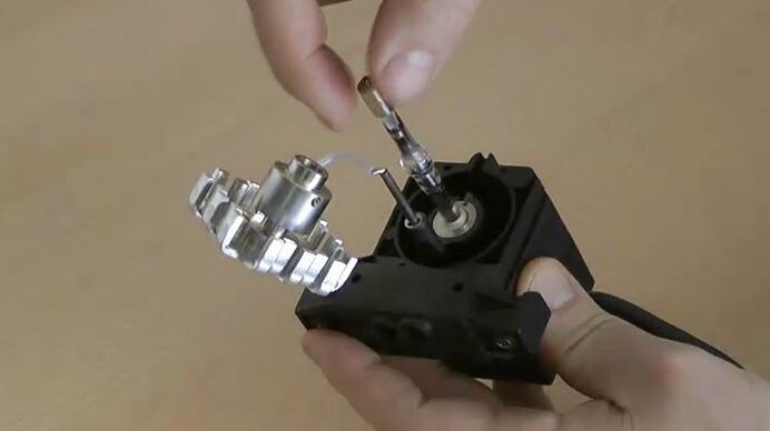
Exchange a fluorescence bulb.

Video Tutorial: How to Change the Bulb of a Fluorescence Lamp Housing

When applying fluorescence microscopy in biological applications, a lamp housing with mercury burner is the most common light source. This video tutorial shows how to change the bulb of a traditional…
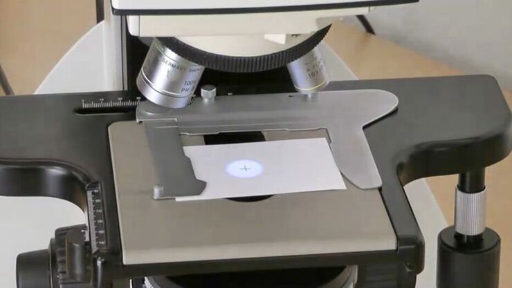
Center a fluorescence bulb.

Video Tutorial: How to Align the Bulb of a Fluorescence Lamp Housing

The traditional light source for fluorescence excitation is a fluorescence lamp housing with mercury burner. A prerequisite for achieving bright and homogeneous excitation is the correct centering and…
Scroll to top