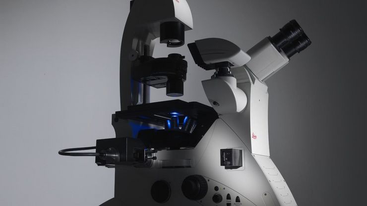
Factors to Consider When Selecting a Research Microscope
An optical microscope is often one of the central devices in a life-science research lab. It can be used for various applications which shed light on many scientific questions. Thereby the…
Focus on Long-Term Imaging in 3D with Light Sheet Microscopy
Long-term 3D imaging reveals how complex multicellular systems grow and develop and how cells move and interact over time, unlocking critical insights into development, disease, and regeneration.…
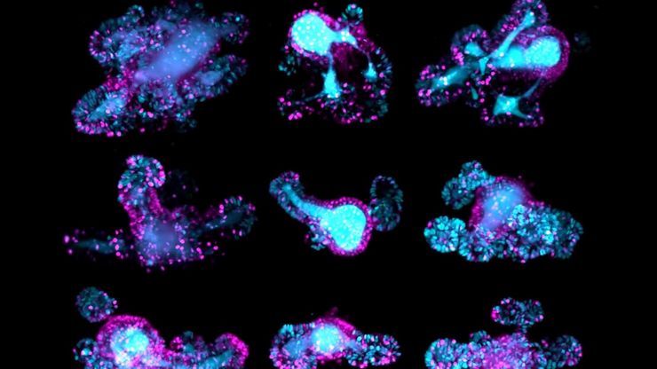
Faster & Deeper Insights into Organoid and Spheroid Models
Gain deeper, more translatable, insights into organoid and spheroid models for drug discovery and disease research by overcoming key imaging challenges. In this eBook, explore advanced microscopy…
A Novel Laser-Based Method for Studying Optic Nerve Regeneration
Optic nerve regeneration is a major challenge in neurobiology due to the limited self-repair capacity of the mammalian central nervous system (CNS) and the inconsistency of traditional injury models.…
How to Image Axon Regeneration in Deep Muscle Tissue
This study highlights Dr. Aaron Lee’s research on mapping nerve regeneration in muscle grafts post-amputation. Limb loss often leads to reduced quality of life, not only from tissue loss but also due…
Capturing Developmental Dynamics in 3D
This application note showcases how the Viventis Deep dual-view light sheet microscope was successfully used by researchers for exploring high-resolution, long-term imaging of 3D multicellular models…
Development and Derisking of CRISPR Therapies for Rare Diseases
This on-demand presentation by Dr. Fyodor Urnov and Dr. Sadik Kassim, originally delivered at ASGCT 2025, focused on a critical challenge in genetic medicine: how to scale CRISPR therapies from…
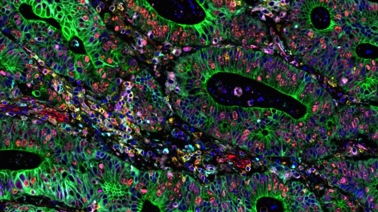
Multiplexed Imaging Reveals Tumor Immune Landscape in Colon Cancer
Cancer immunotherapy benefits few due to resistance and relapse, and combinatorial therapeutic strategies that target multiple steps of the cancer-immunity cycle may improve outcomes. This study shows…
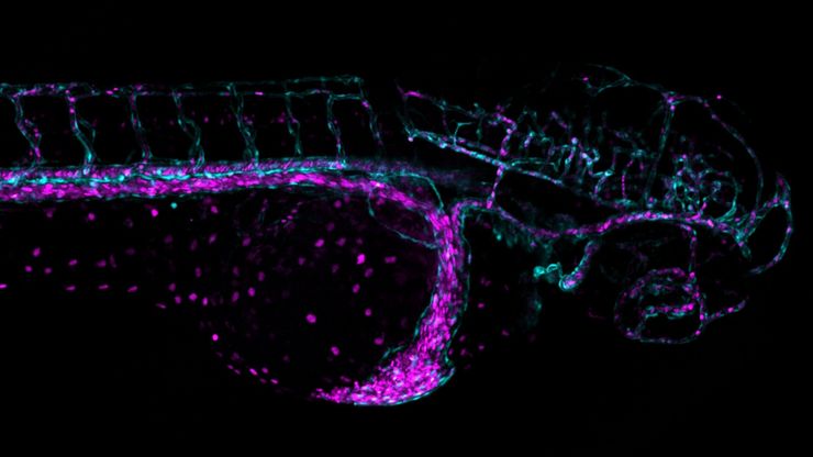
Overcoming Challenges with Microscopy when Imaging Moving Zebrafish Larvae
Zebrafish is a valuable model organism with many beneficial traits. However, imaging a full organism poses challenges as it is not stationary. Here, this case study shows how zebrafish larvae can be…


