
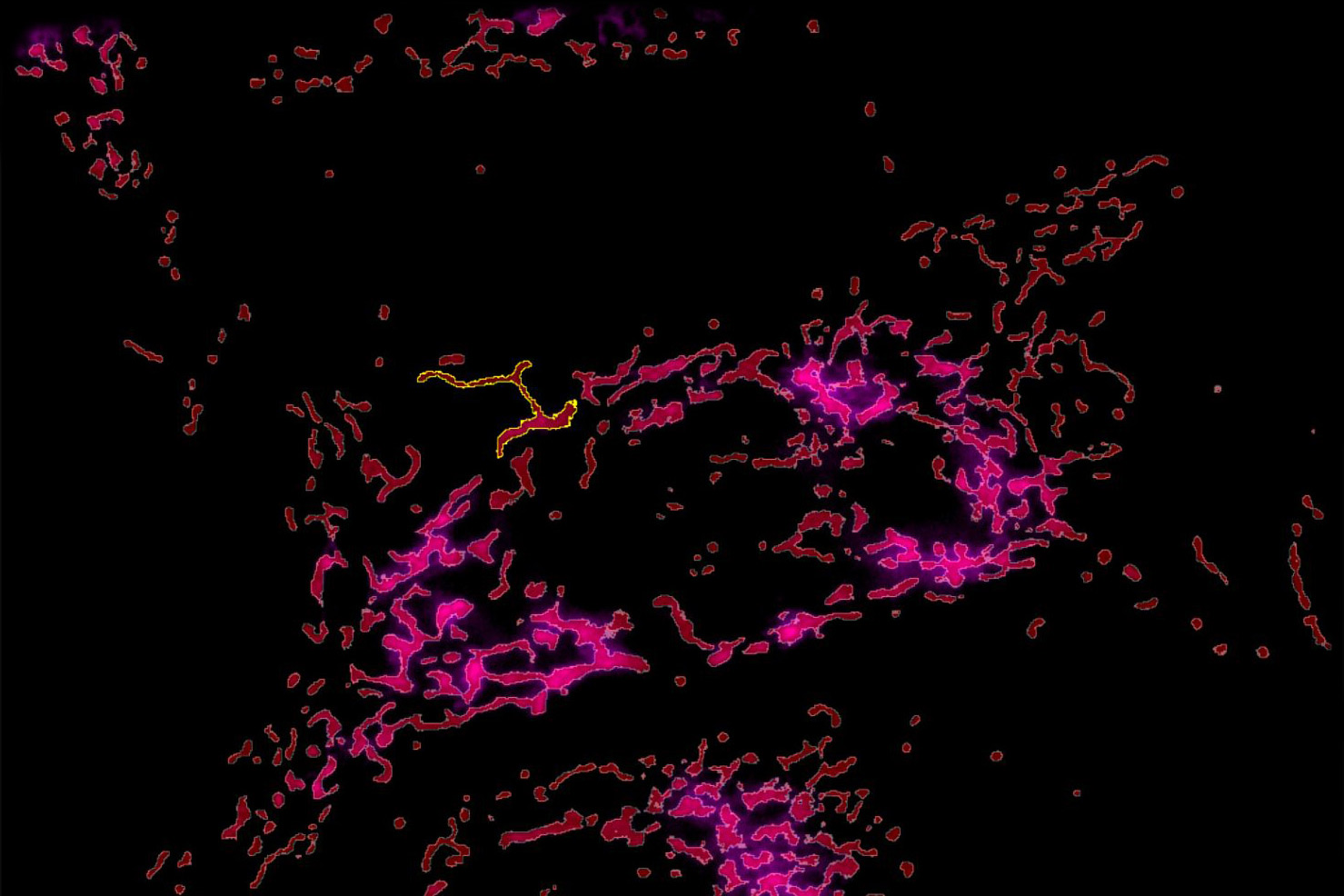
U2OS cells stained with MitoTracker green (Mitochondria structure, cyan) and TMRE (active mitochondria, magenta). Sequential acquisition of the two channels over 2 minutes 100 frames using the 63x/1.20 CS2 Water MotCORR objective.
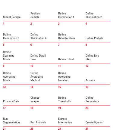
Spheroid Growth over 2.5 days
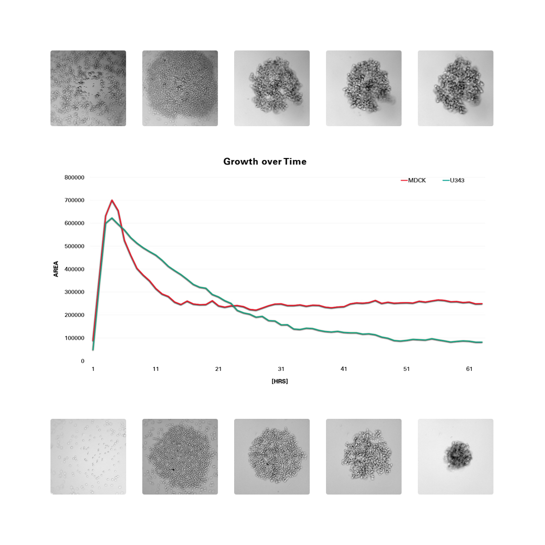
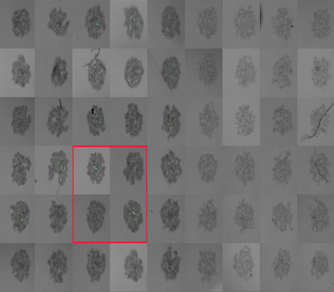
Formation of 3D spheroids from 1000 stably transfected MDCK MX1-GFP cells per well (upper row) and 1000 U2OS cells per well (lower row). Time-lapse acquisition over 60 hrs with 30 minutes interval. Green, GFP. Black and white, integrated modulation contrast.
Formation of 3D spheroids from 1000 stably transfected MDCK MX1-GFP cells per well (left half) and 1000 U2OS cells per well (right half) shown at 5 different timepoints. Time -lapse acquisition over 60 hrs. with 30 minutes interval. Green, GFP. Gray, integrated modulation contrast.
Mount sample
Mount sample
Position sample
Position sample
Define illumination 1
Start Live Acquisition
Define illumination 2
Define positions
Define illumination 3
Acquire
Define illumination 4
Choose images
Define detector gain
Run segmentation
Define pinhole
Run analysis
Define scanning mode
Define dwell time
Define offset
Define line step
Define averaging mode
Define averaging method
Define averaging number
Acquire
Process data
Choose images
Define thresholds
Define separators
Run segmentation
Run analysis
Extract information
Create figures


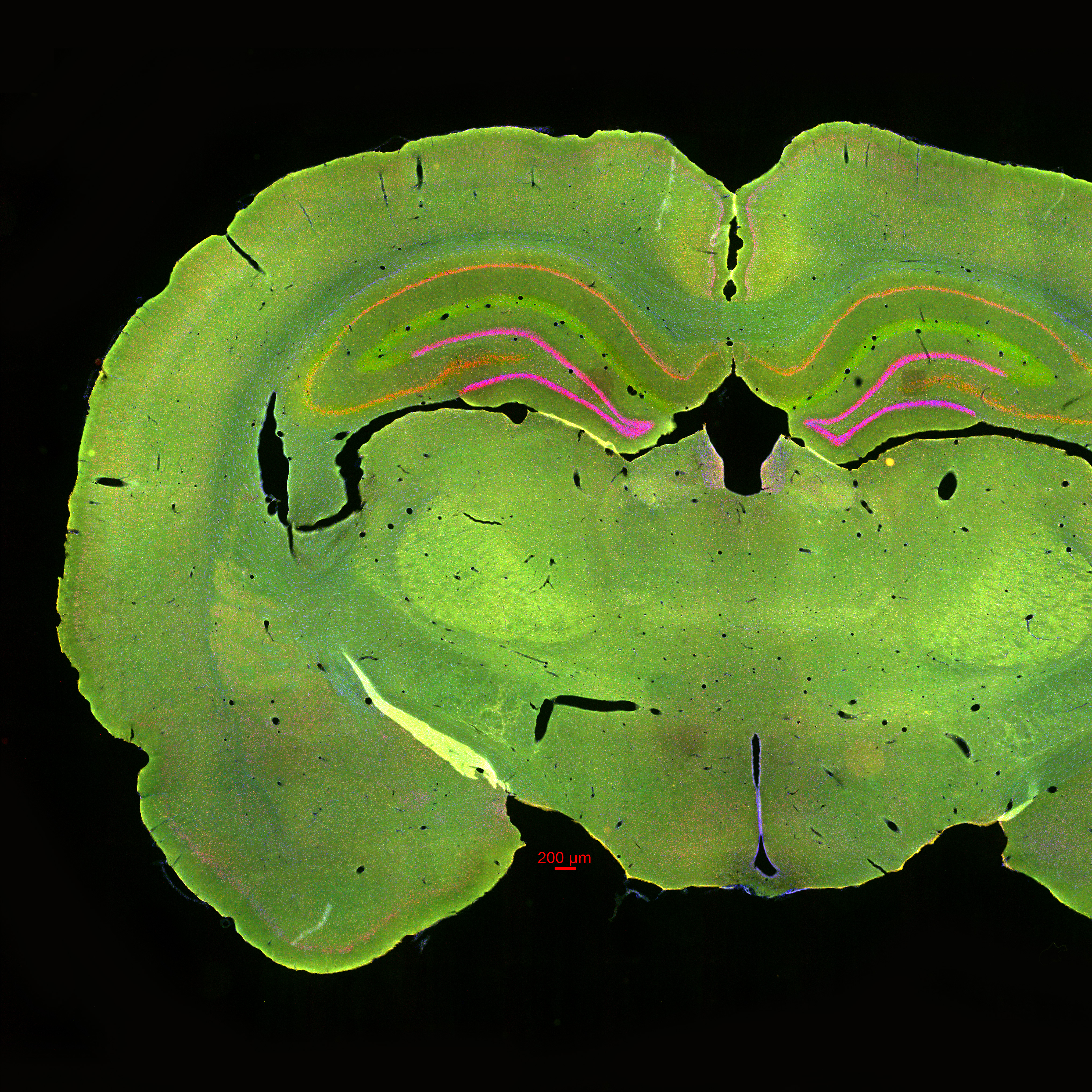
Tissue slice from the rat brain. Nuclei are stained with DAPI (blue), STL with FITC (green), astrocytes (GFAP) with Cy3 (yellow), and newborn neurons (NeuN) with Cy5 (red). 10x widefield tile scan, all 4 labels aquired simultaneously.
U2OS cells stained with MitoTracker green (Mitochondria structure, cyan) and TMRE (active mitochondria, magenta). Simultaneous acquisition of the two channels over 2 minutes 100 frames using the 63x/1.20 CS2 Water MotCORR objective.
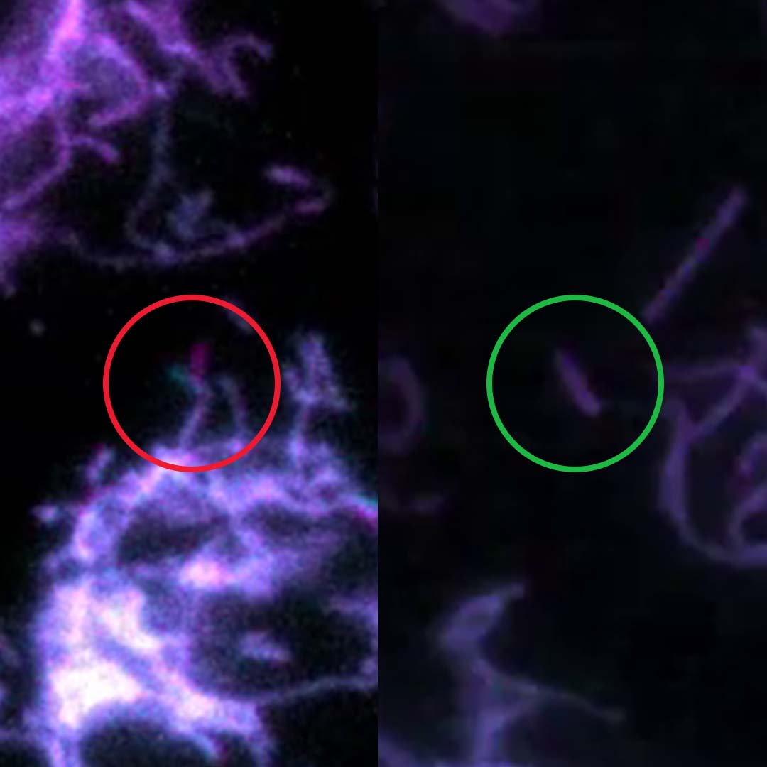
Sequential acquisition with a conventional microscope
Simultaneous acquisition with Mica
U2OS cells stained with MitoTracker green (Mitochondria structure, cyan) and TMRE (active mitochondria, magenta). Simultaneous acquisition of the two channels over 2 minutes 100 frames using the 63x/1.20 CS2 Water MotCORR objective.
3D Cell Culture, 7d spheroid formation of U343 cells. tfLC3 EGFP and mRFP + DAPI + WGA Alexa680. Objective: 20x/0.75 CS2 DRY
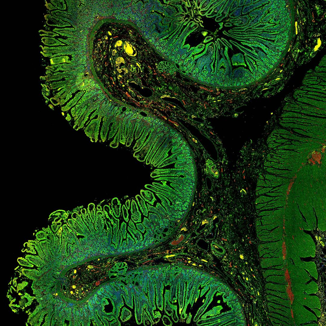
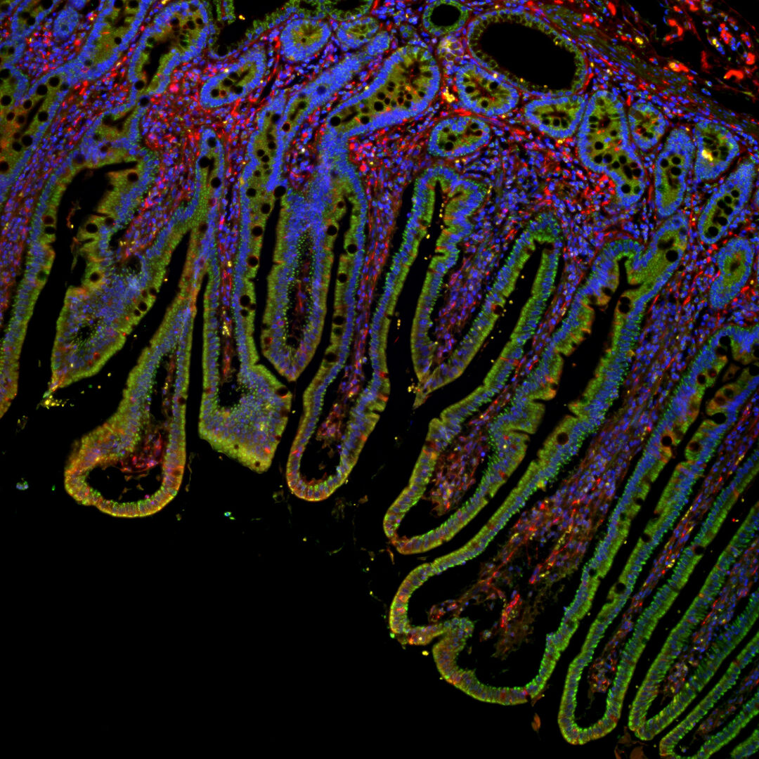
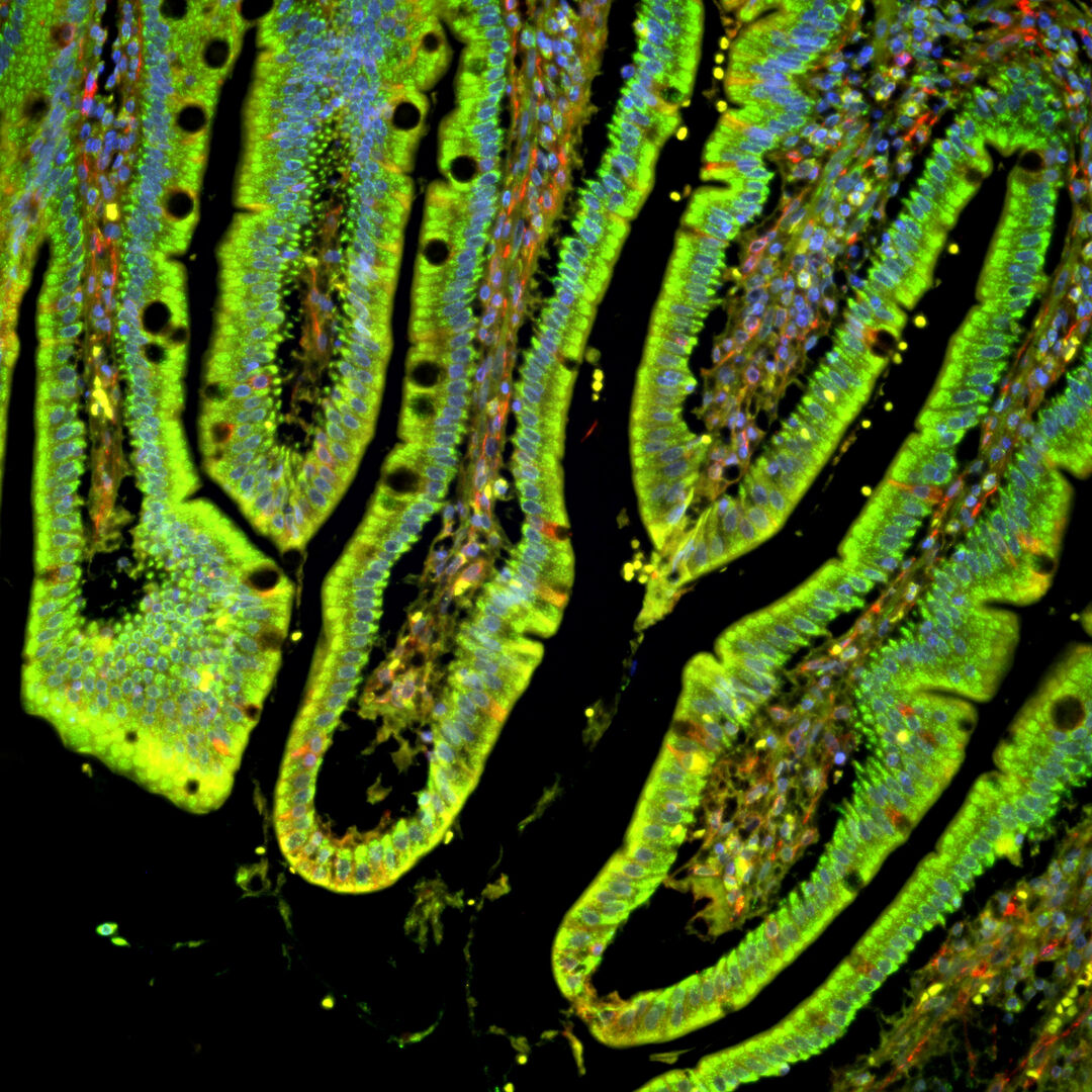
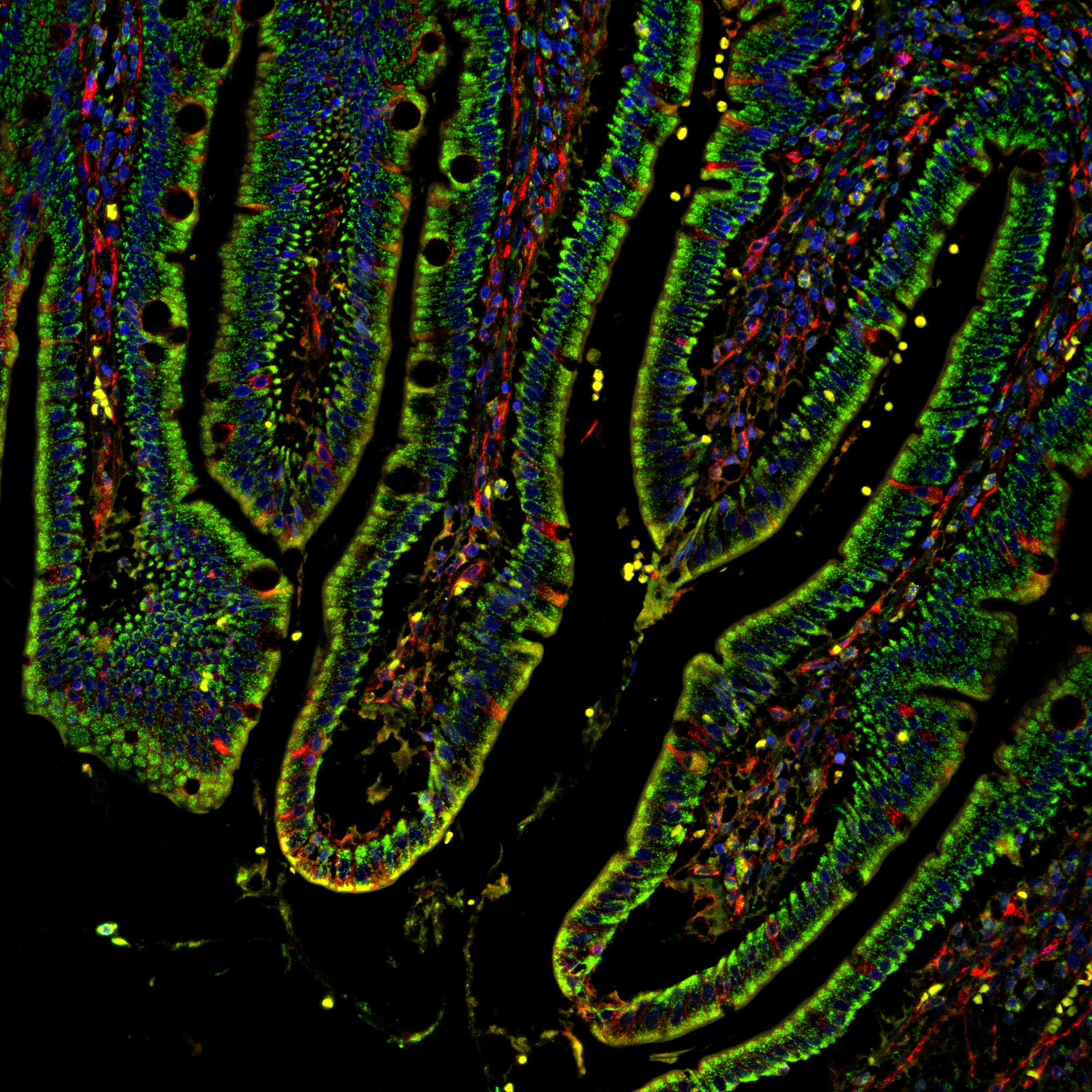
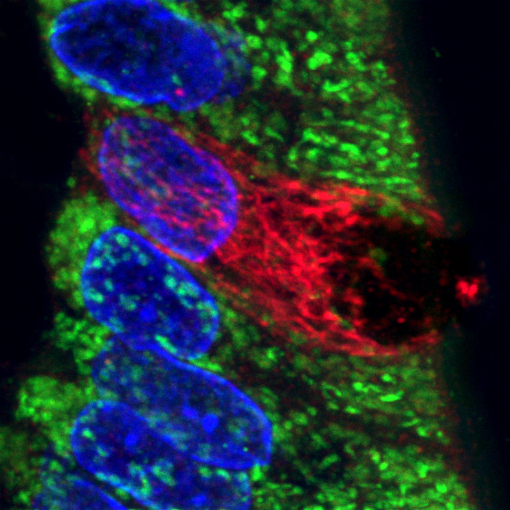
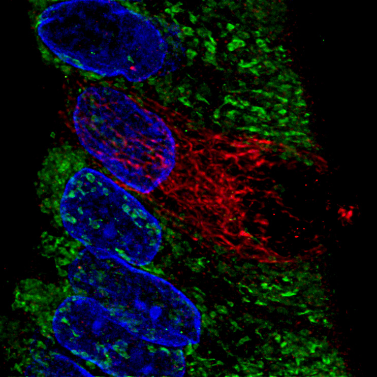
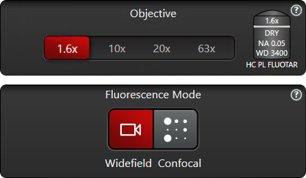
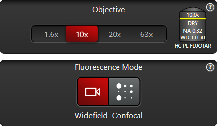
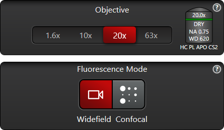
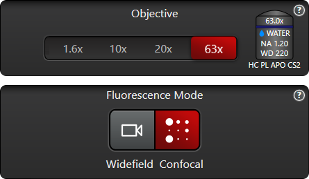
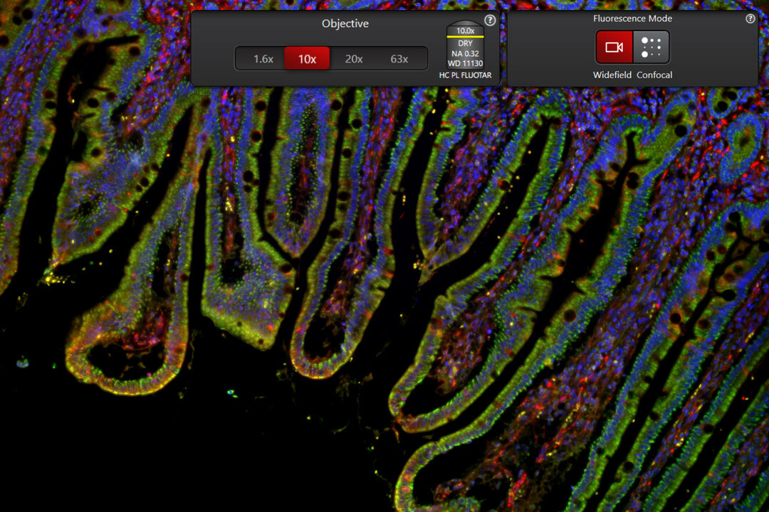
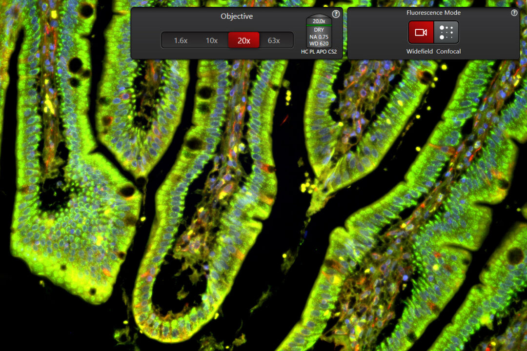
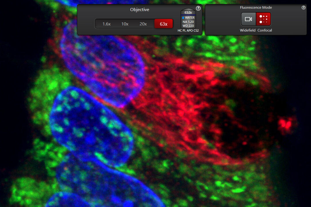
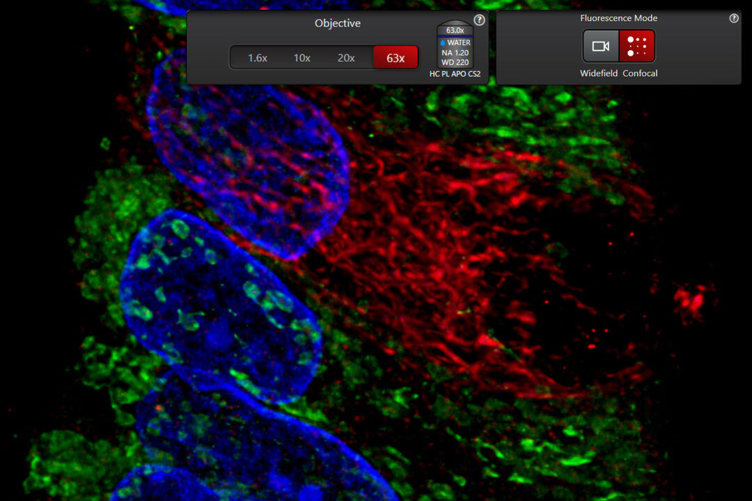
Intestine tissue section acquired with different objectives ranging from low to high magnification (1.6x, 10x, 20x, 63x), using widefield and confocal imaging. 20x widefield images are processed with THUNDER and 63x confocal images with LIGHTNING. Nuclei are labeled in blue, mitochondria in green, and detyrosinated tubulin in red.

U2OS cells were labelled with SiR-Actin, TMRE (mitochondria activity), CellEvent™ (caspase activity), and DAPI (nuclei). Apoptosis inducer staurosporine was added at time-point 0. 63x magnification, widefield mode. 13 hours time-lapse.

Eliminate over 85% of tedious setup steps that require special expertise
Step into the era of Access for all
Everyone can now leverage microscopy to make more discoveries
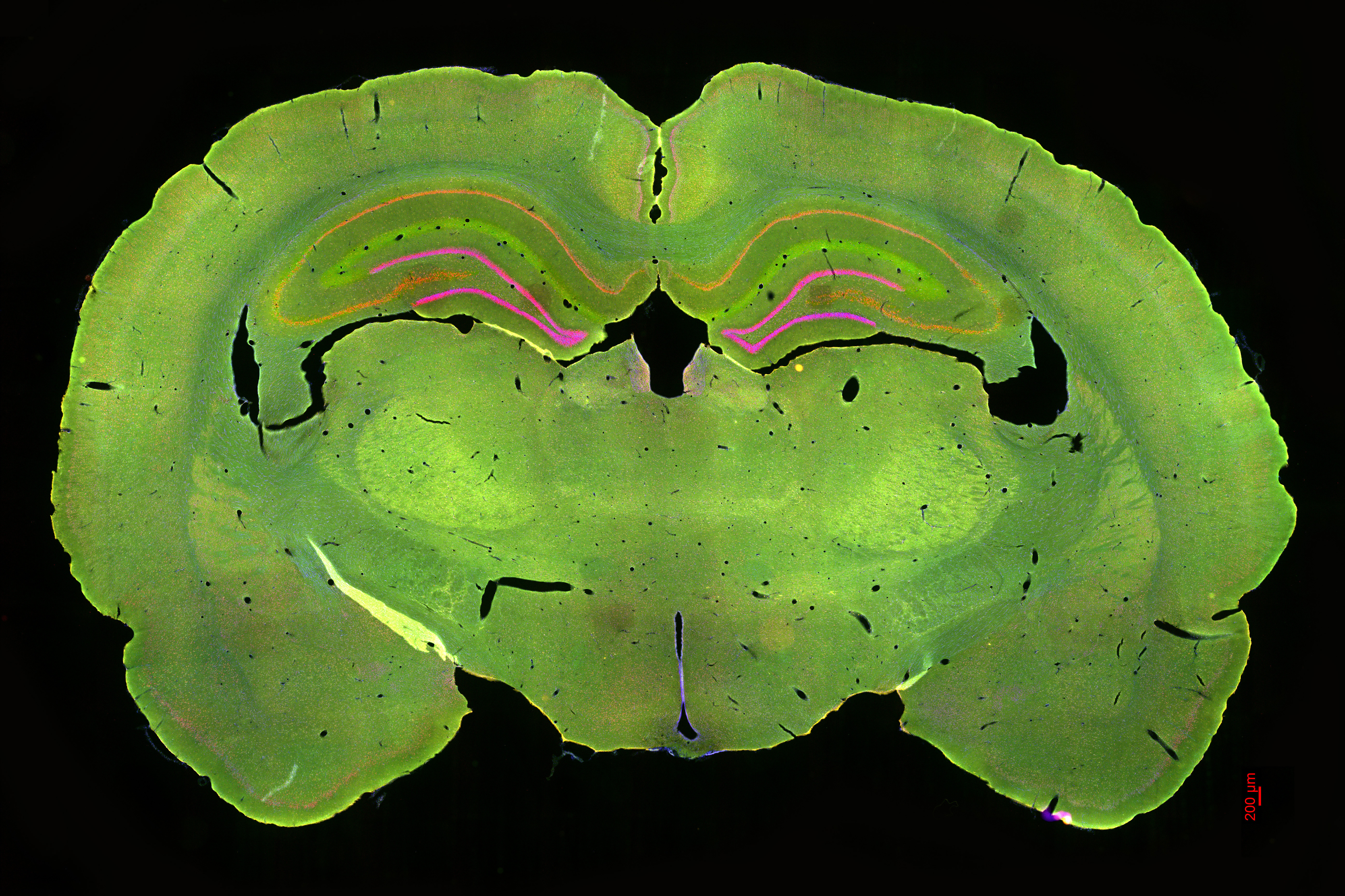
Tissue slice from the rat brain. Nuclei are stained with DAPI (blue), STL with FITC (green), astrocytes (GFAP) with Cy3 (yellow), and newborn neurons (NeuN) with Cy5 (red). 10x widefield tile scan, all 4 labels aquired simultaneously.
85% fewer steps to the first image
1/3 less time to the first image
1/2 of the training time
Powered by:
Intelligent automation
Intelligent imaging

Step into the era of No constraints
The Microhub: everything you need to enable discoveries, unified in one easy-to-use system
4x more data with
100% correlation
Access key contextual information with absolute spatiotemporal correlation
U2OS cells stained with MitoTracker green (Mitochondria structure, cyan) and TMRE (active mitochondria, magenta). Sequential acquisition of the two channels over 2 minutes 100 frames using the 63x/1.20 CS2 Water MotCORR objective.
Sequential acquisition with a conventional microscope
U2OS cells stained with MitoTracker green (Mitochondria structure, cyan) and TMRE (active mitochondria, magenta). Simultaneous acquisition of the two channels over 2 minutes 100 frames using the 63x/1.20 CS2 Water MotCORR objective.
Simultaneous acquisition with Mica
Mica delivers absolute correlated labels without spatiotemporal mismatch
Powered by:
4 labels simultaneously
4 labels 100% correlated
Patented FluoSync technology
Seamlessly move from fast overview to high resolution when required by your experiment
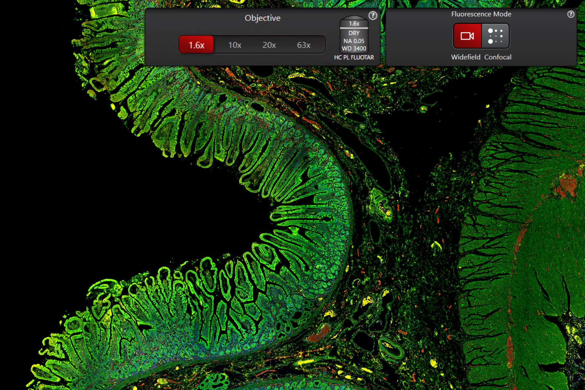
Intestine tissue section acquired with different objectives ranging from low to high magnification (1.6x, 10x, 20x, 63x), using widefield and confocal imaging. 20x widefield images are processed with THUNDER and 63x confocal images with LIGHTNING. Nuclei are labeled in blue, mitochondria in green, and detyrosinated tubulin in red.
Create Overview
Find the sample structure on the carrier and observe the overall morphology of the colon slice. Identify a region of interest for more detailed inspection.
Get more details of a substructure
Switching to the next higher magnification allows to assess the integrity of the tissue and locate areas suitable for further analysis.
Select the cell of interest
Start to see the higher details and select the single cell to get subcellular information. However, some details remain hidden in the haze.

Select the cell of interest
THUNDER is the method of choice to get more contrast and see more details. This enables you to make the right selection and step further into the details of the sample.
Get the subcellular information
Switch from Widefield to Confocal mode with just a simple click to get more subcellular information.
Get even more of the subcellular information
Adding LIGHTNING gives access to higher details of the subcellular structures seamlessly integrated into the whole workflow from fast overview to high resolution.
Powered by:
Unified imaging modalities
Point scanning confocal
Mica is an incubator
Formation of 3D spheroids from 1000 stably transfected MDCK MX1-GFP cells per well (upper row) and 1000 U2OS cells per well (lower row). Time-lapse acquisition over 60 hrs with 30 minutes interval. Green, GFP. Black and white, integrated modulation contrast.
Achieve physiological-like conditions throughout your experiment
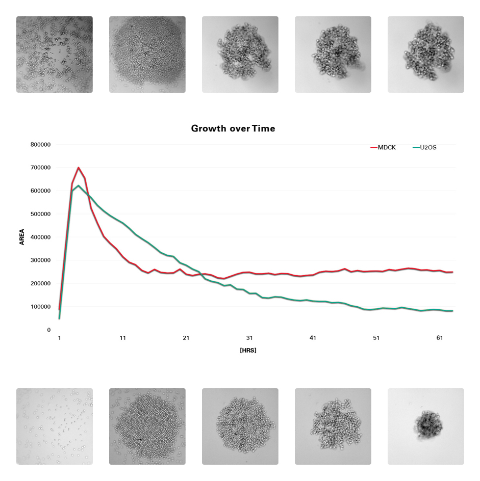
Formation of 3D spheroids from 1000 stably transfected MDCK MX1-GFP cells per well (left half) and 1000 U2OS cells per well (right half) shown at 5 different timepoints. Time -lapse acquisition over 60 hrs. with 30 minutes interval. Green, GFP. Gray, integrated modulation contrast.
Mica is an incubator to maintain your sample in optimal conditions and to minimize evaporation

Step into the era of Radically simplified workflows
Bringing you faster from sample to discovery
Reduce over 60% of process steps through system intelligence
Conventional microscopes
With conventional microscopes, you need to define various steps, from sample to analysis, that you need to run when you set up an experiment.
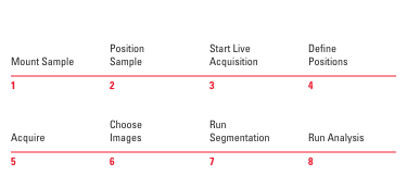
Mica automation
With Mica, you can reduce time and effort by radically simplifying your workflow into as few as 8 steps from sample to insight through system intelligence.
Powered by:
Sample Finder
OneTouch-Auto-Illumination
AI-based analysis
Reduce time and effort from sample to insight by simplifying your entire workflow
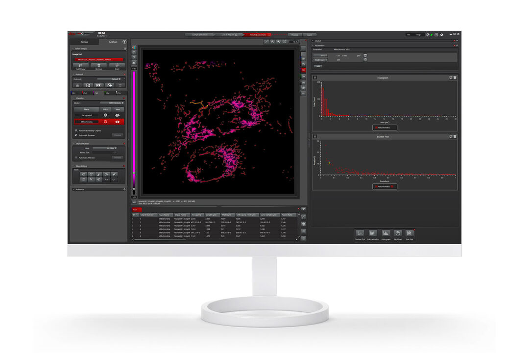
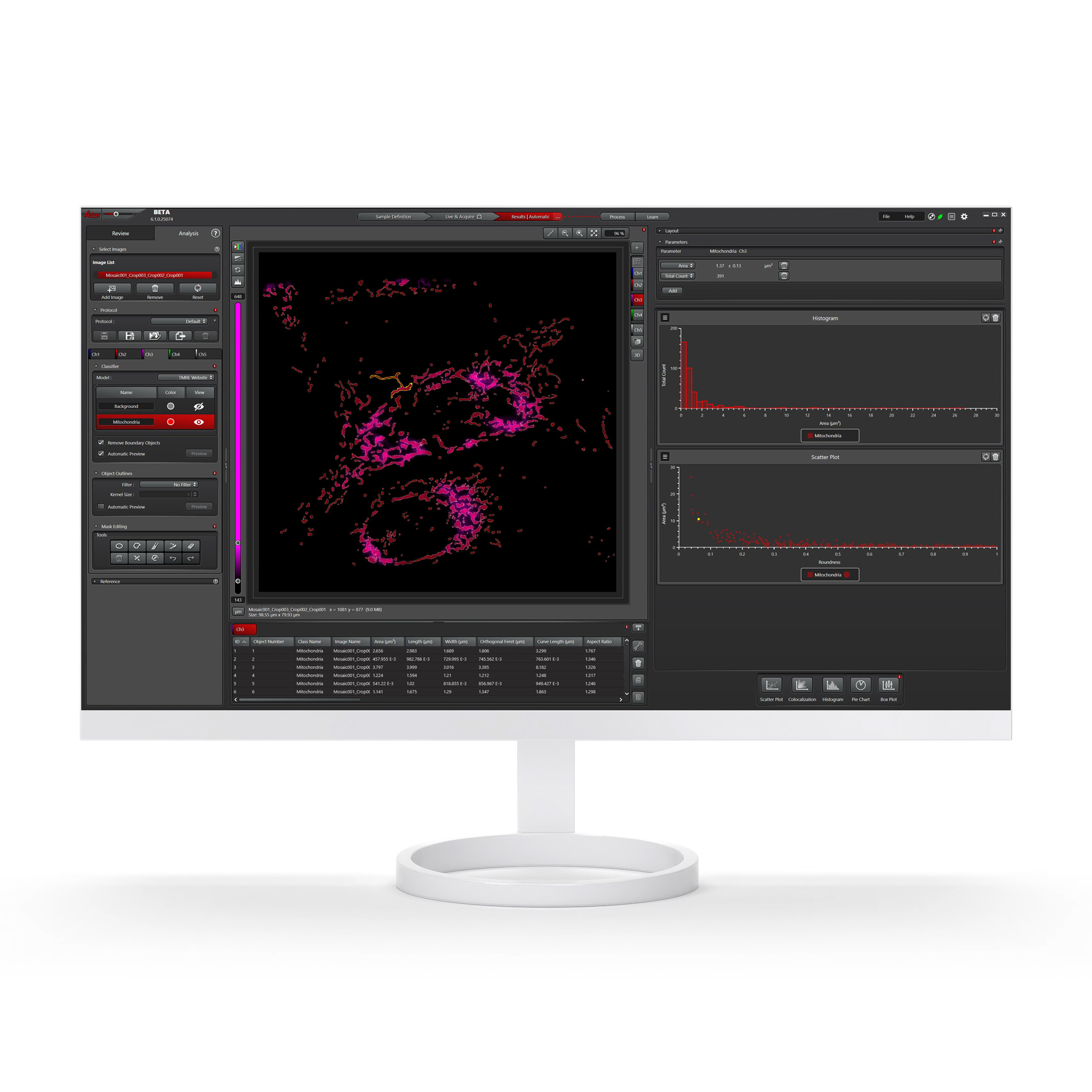
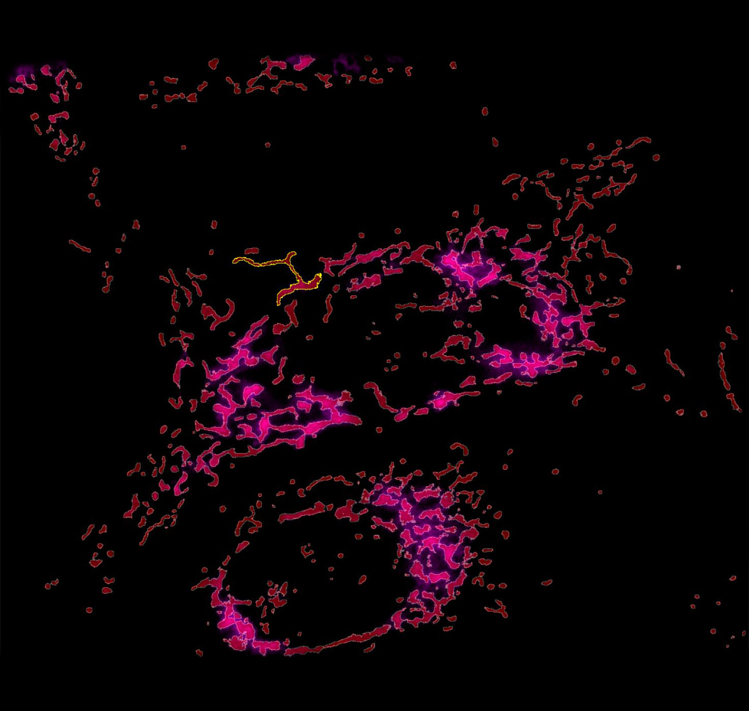
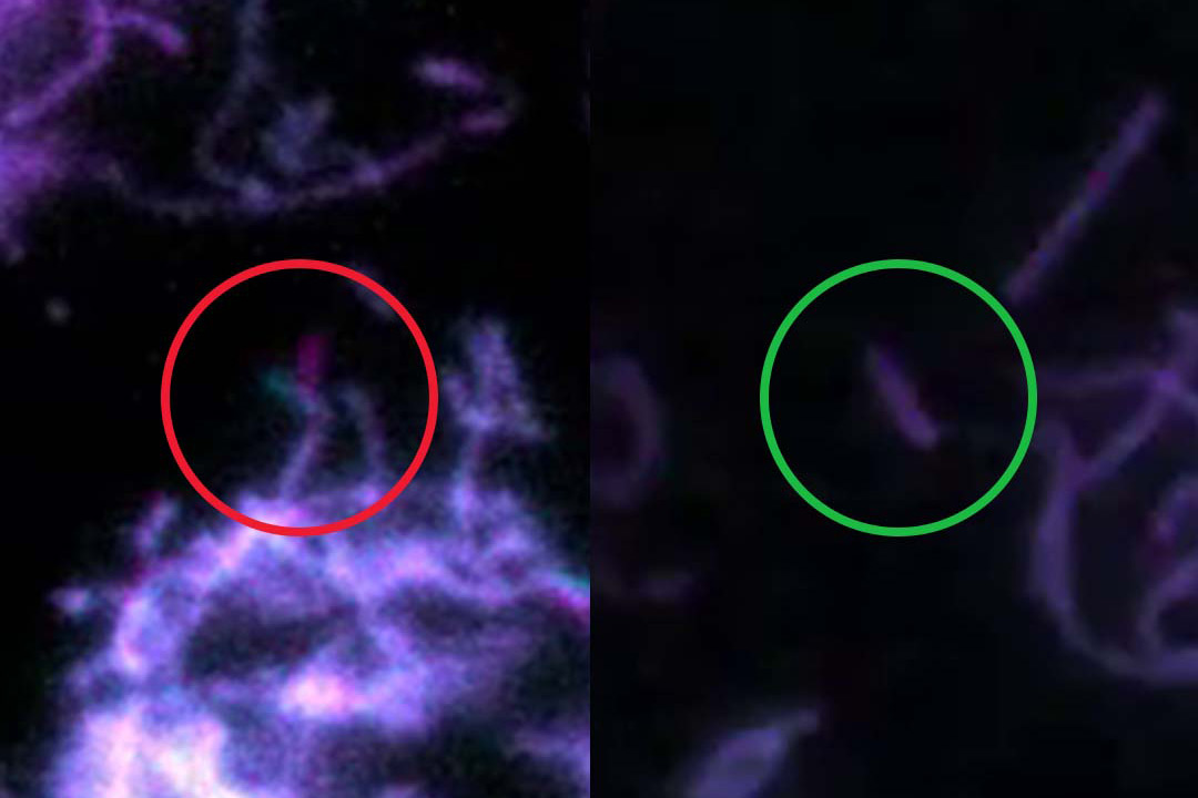
AI based training of mitochondrial segmentation using your scientific expertise
U2OS cells were labelled with SiR-Actin, TMRE (mitochondria activity), CellEvent™ (caspase activity), and DAPI (nuclei). Apoptosis inducer staurosporine was added at time-point 0. 63x magnification, widefield mode. 13 hours time-lapse.
Enable 100% reproducibility and repeatability throughout your experiment
Powered by:
Pixel classifier
GUI operated annotations
Reusable AI models and projects parameters
Meet Mica
The Microhub era is now! Experience the future.
Meet Mica in key applications
Fluorescence multi-well plate Assay
Mica allows you to image 4 labels simultaneously, with 100% spatiotemporal correlation. This key application shows how Mica is used with fluorescent multi-well plate assays around Caspase 3/7 activations in apoptosis.
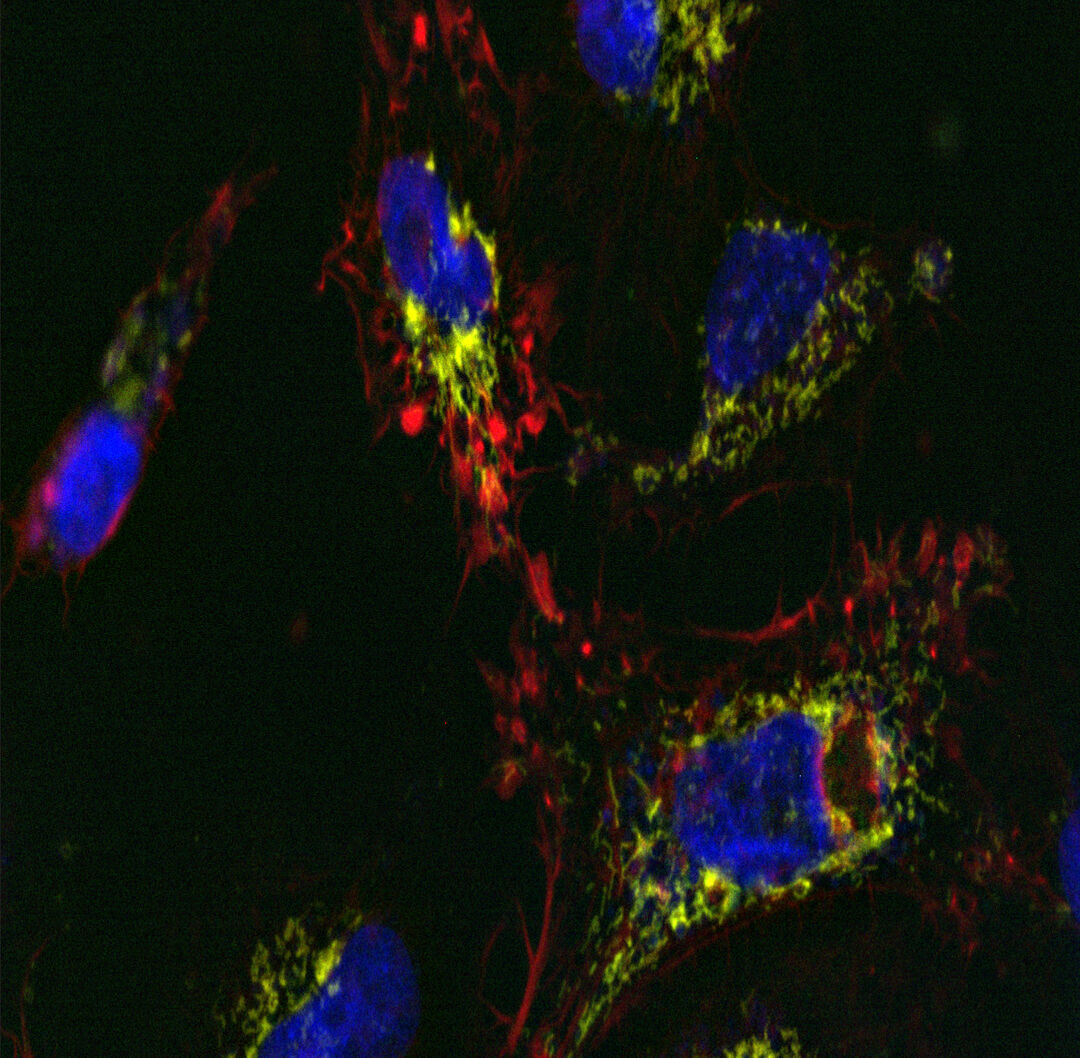
U2OS cells were labelled with SiR-Actin, TMRE (mitochondria activity), CellEvent™ (caspase activity), and DAPI (nuclei). Apoptosis inducer staurosporine (3µM) was added at time-point 0. 63x magnification, widefield mode. 13 hours time-lapse.
3D Tissue Imaging
Mica enables you to seamlessly move from fast overview to high resolution when required by your experiment. See how Mica allows you identify a detyrosynated tubulin positive cell and progress from overview to the segmentation of the tubulin network.
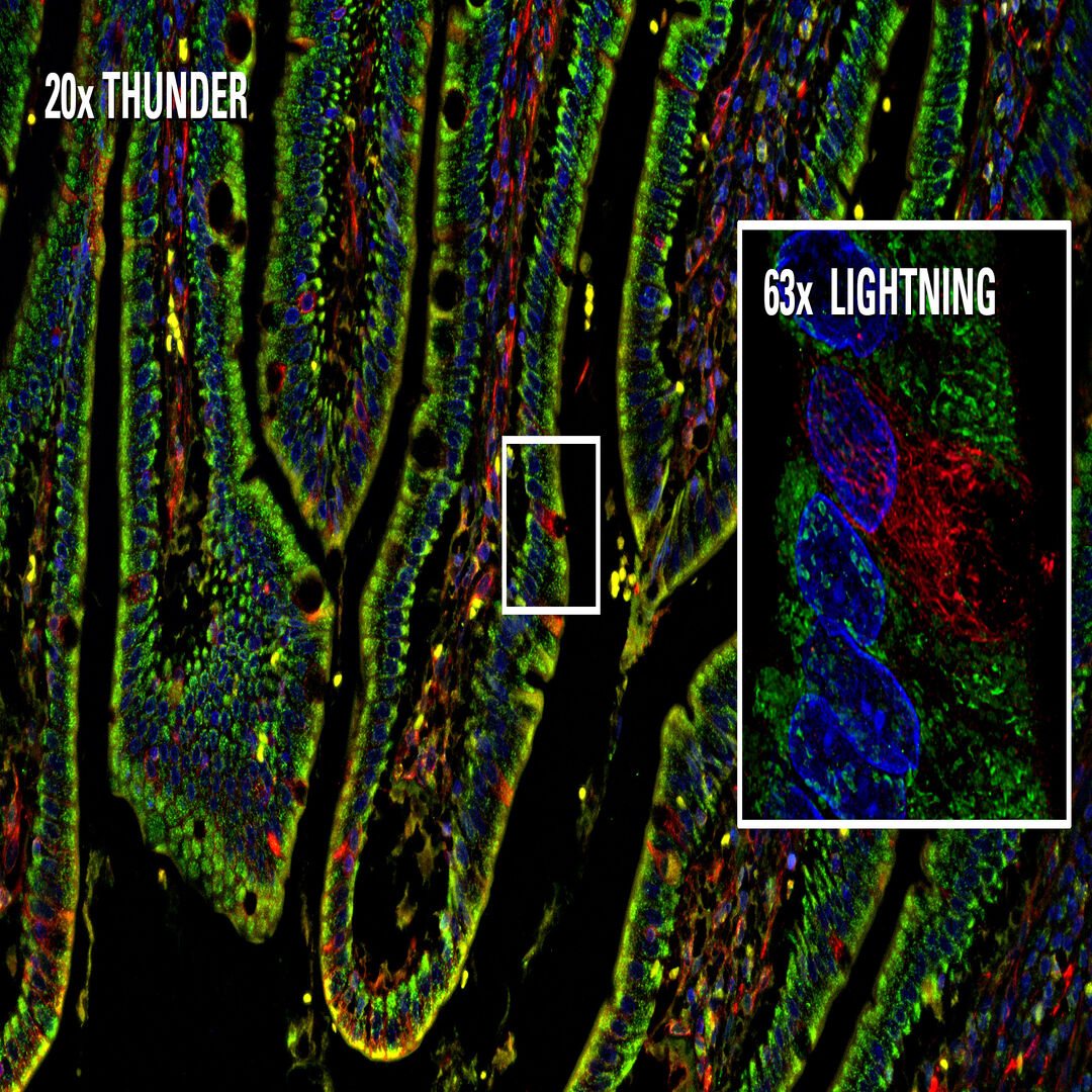
Intestine tissue section acquired with 20x and 63x magnification, using widefield and confocal imaging. 20x widefield images are processed with THUNDER and 63x confocal images with LIGHTNING. Nuclei are labeled in blue, mitochondria in green, and detyrosynated tubulin in red.
Long-term Time lapse
Mica is an incubator to maintain your sample in physiological-like conditions and to minimize evaporation. Discover how Mica allows you to measure spheroids growth and to analyze protein expression levels.
Formation of 3D spheroids from 1000 stably transfected MDCK MX1-GFP cells per well and 1000 U2OS cells per well shown at 5 different timepoints. Time-lapse acquisition over 60 hrs. with 30 minutes interval. Green, GFP. Gray, integrated modulation contrast.

What if every scientist could access spatial information?
Popular Configurations

Mica 20x LWD Objective with Service Installation20x long working distance objective for imaging on glass and plastic bottom carriersTo penetrate deeper into samples using everyday standard plastic-bottom multi well plates, this Long Working Distance (LWD) objective has been optimized for use with Mica.
Why is long working distance important?
Please note: This objective requires service installation to ensure correct installation and calibration. A half-day service visit by our expert team is added to ensure optimal performance and system compatibility.
Loading...
View product details+
Product includes
Loading...
|
||||||||||||||||||||||||||||||||||||

Mica Widefield Live Cell Microhub Automated MicroscopeSimplify advanced live cell and fluorescence imaging. Get reliable, expert level microscopy results faster with extensive intelligent automation, AI-supported analysis, and an interface designed for ease of use. Perform long-term live cell experiments up to 4x faster than traditional widefield systems, capturing events that may previously have been missed. Easily get quality images as Mica automatically corrects the focus during experiments. Minimise phototoxicity and maintain optimal cell health with full environmental control. Ideal for long-term live-cell studies at near physiological conditions. MICA BENEFITS: ACCESS FOR ALL: Mica requires minimal technical input and gives consistent results every time, so all lab members can leverage microscopy to make more discoveries. NO CONSTRAINTS: Get true simultaneous capture of up to four labels without spatiotemporal mismatch – at near-physiological conditions for live cells, or with fixed samples. RADICALLY SIMPLIFIED WORKFLOWS: Intelligent automation and AI-supported analysis of diverse samples, such as tissues on slides or well plates, enable greater efficiency and a faster track to publication.
Loading...
View product details+
Product includes
Loading...
|
||||||||||||||||||||||||||||||||||||
Mica Widefield Microhub Automated MicroscopeSimplify advanced fluorescence imaging through extensive intelligent automation, AI‑supported analysis, and an easy-to-use interface. Now all lab members can get expert microscopy results, regardless of experience. Ideal for exploration and high-throughput fluorescence imaging of fixed samples. MICA BENEFITS: ACCESS FOR ALL: Mica requires minimal technical input and gives consistent results every time, so all lab members can leverage microscopy to make more discoveries. NO CONSTRAINTS: Get true simultaneous capture of up to four labels for fast acquisitions. RADICALLY SIMPLIFIED WORKFLOWS: Intelligent automation and AI-supported analysis of diverse samples, such as tissues on slides or well plates, enable greater efficiency and a faster track to publication.
Loading...
View product details+
Product includes
Loading...
|
||||||||||||||||||||||||||||||||||||
Mica WideFocal Live Cell Microhub Automated MicroscopeCombine widefield and confocal imaging in an incubating environment and simplify advanced live cell and fluorescence imaging. Extensive intelligent automation, AI-supported analysis, and an easy-to-use interface help you achieve expert microscopy results without all the training. Perform long-term live cell experiments up to 4x faster than traditional widefield systems, capturing events that may previously have been missed. Easily get quality images as Mica automatically corrects the focus during experiments. Minimise phototoxicity and maintain optimal cell health with full environmental control. Ideal for long-term and dynamic live cell studies of organoids, spheroids, and 3D cultures. MICA BENEFITS: ACCESS FOR ALL: Mica requires minimal technical input and gives consistent results every time, so all lab members can leverage microscopy to make more discoveries. NO CONSTRAINTS: Visualize in widefield and switch effortlessly to confocal without ever moving your sample. Get true simultaneous capture of up to four labels without spatiotemporal mismatch – at near-physiological conditions for live cells, or with fixed samples. RADICALLY SIMPLIFIED WORKFLOWS: Intelligent automation and AI-supported analysis of diverse samples, such as tissues on slides or well plates, enable greater efficiency and a faster track to publication.
Loading...
View product details+
Product includes
Loading...
|
||||||||||||||||||||||||||||||||||||
Mica WideFocal Microhub Automated MicroscopeCombine widefield and confocal imaging in an easy-to-use platform and simplify advanced fluorescence imaging. By streamlining workflows and reducing complexity through intelligent automation, AI-supported analysis, you can get expert microscopy results without all the training. Ideal for high-resolution imaging of thick or complex samples. MICA BENEFITS: ACCESS FOR ALL: Mica requires minimal technical input and gives consistent results every time, so all lab members can leverage microscopy to make more discoveries. NO CONSTRAINTS: Visualize in widefield and switch effortlessly to confocal without ever moving your sample. Get true simultaneous capture of up to four labels for fast acquisitions. RADICALLY SIMPLIFIED WORKFLOWS: Intelligent automation and AI-supported analysis of diverse samples, such as tissues on slides or well plates, enable greater efficiency and a faster track to publication.
Loading...
View product details+
Product includes
Loading...
|
||||||||||||||||||||||||||||||||||||

Don’t see your Configuration? Request for individual Quote
