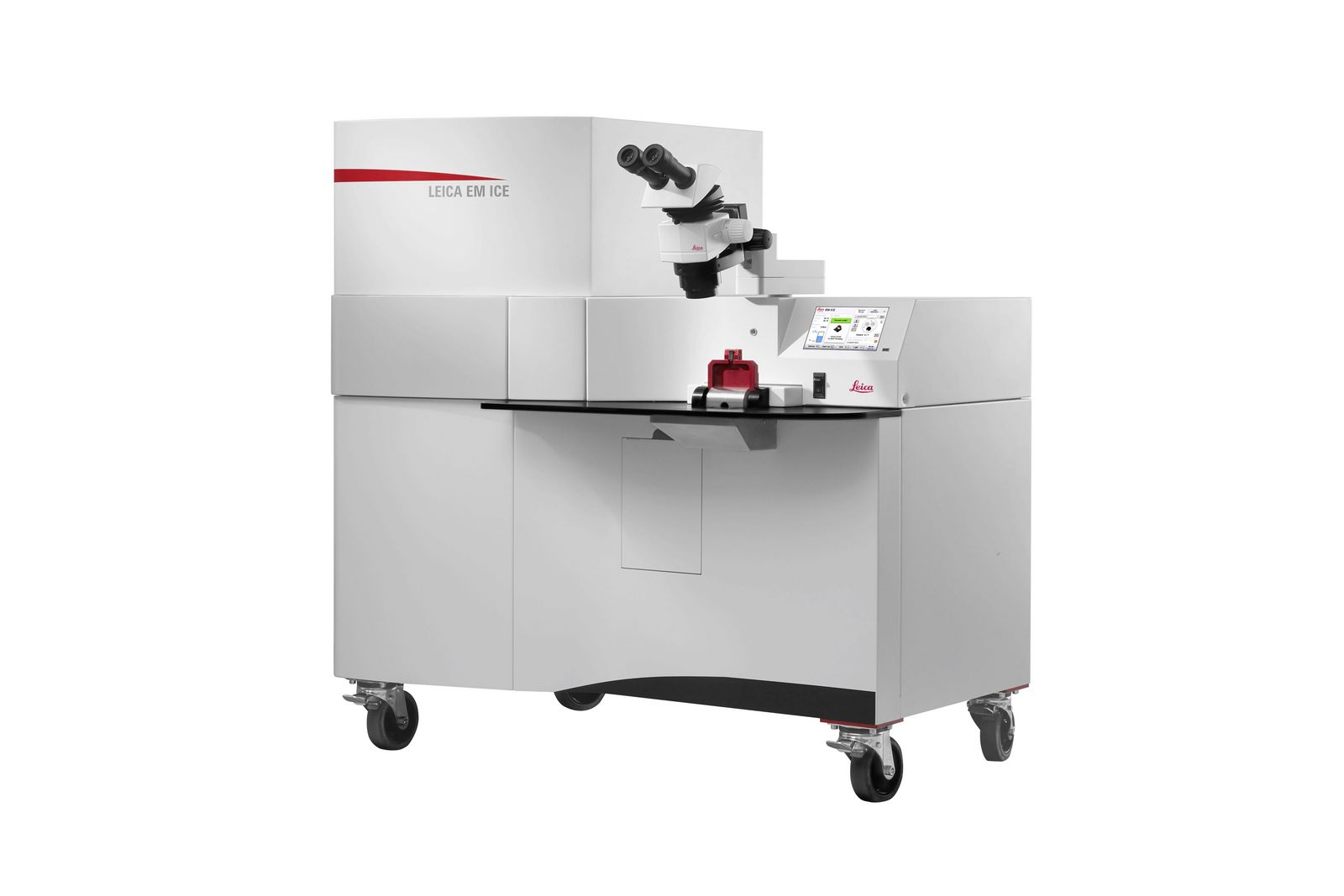
EM ICE Versatile High Pressure Freezing platform
High-pressure freezing is essential for detailed analysis of cellular components, as it enables rapid cryo-immobilization of aqueous samples while preserving their ultrastructure. EM ICE combines advanced high-pressure freezing with light and electrical stimulation, making it the ideal platform for groundbreaking discoveries in electron and correlative microscopy.
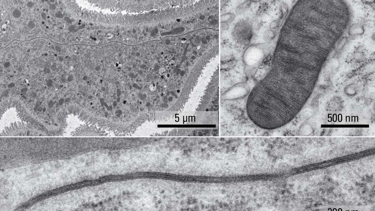
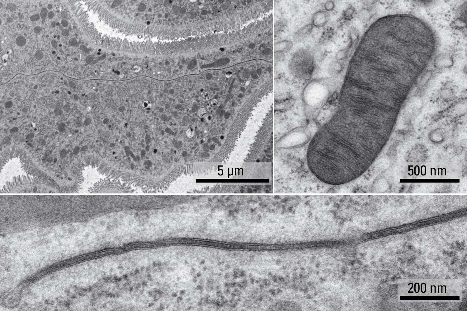
Preserve samples in their native states
Cryo-fixation is the only way to fix cellular constituents without introducing significant structural alterations.
The EM ICE high pressure freezer uses a unique, alcohol-free freezing principle to allow for a superior cryo-fixation of the specimen enabling better quality results to be obtained:
- No alcohol in the chamber leads to a faster pressure increase and immediate cooling of the specimen
- No alcohol residue on the carrier or the specimen
- One move: fully automated loading to freezing in 1 second
Freeze your sample for a range of applications
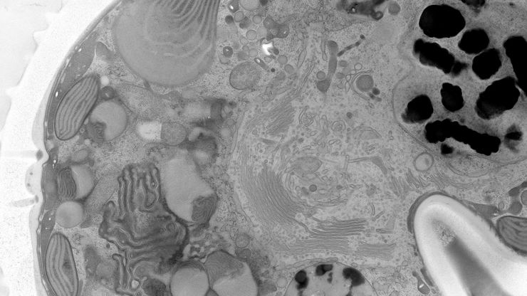
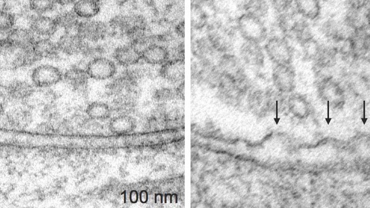
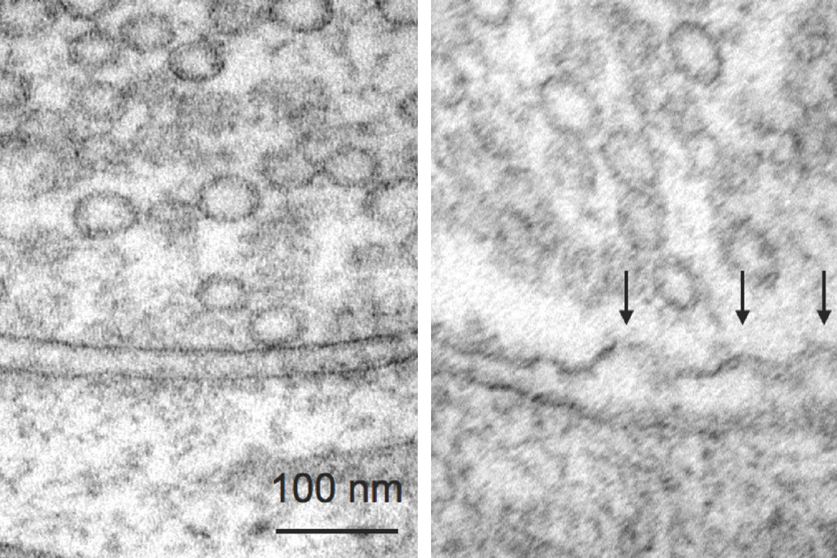
High-quality frozen samples are the essential prerequisite for advanced cryo-EM workflows like cryo-electron tomography (Cryo-ET) and on-grid lamella preparation, including waffle freezing.
Sample structure preservation means the EM studies observation reflects the true physiological state of the sample at an unparallel resolution.
High pressure freezing with EM ICE is a starting point for 3D volume EM workflows like Array Tomography and serial block face imaging. After successful vitrification samples undergo freeze substitution to enable detailed electron microscopy studies.
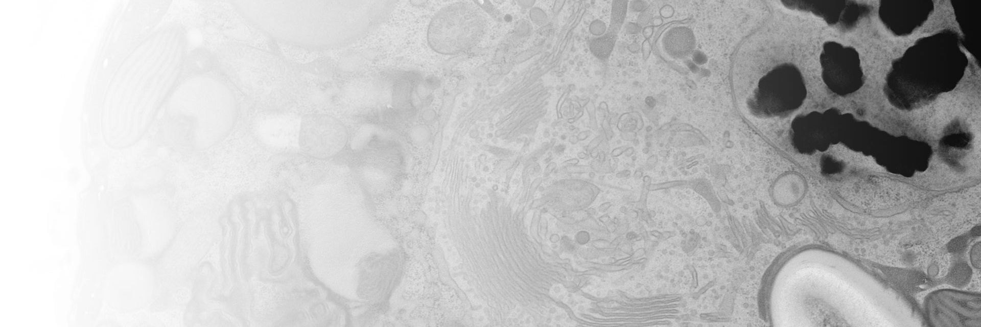
Video: Matsui, Spangler et al. “Cryo-electron tomographic investigation of native hippocampal glutamatergic synapses” https://doi.org/10.7554/eLife.98458.3.sa0
https://creativecommons.org/licenses/by/4.0/ - Adapted from Figure 5 video 1
Freeze your sample when the important event happens
Tailor your EM ICE by combining light or electrical stimulation with high-pressure freezing to capture dynamic events.
EM ICE is a reliable solution for capturing action potential and membrane trafficking processes.
- Electrical stimulation with millisecond precision, complete coordination of electrical discharge at the moment of freezing
- Rigorous correlation between the light pulses and the moment of freezing for precise light stimulation of any light sensitive compounds
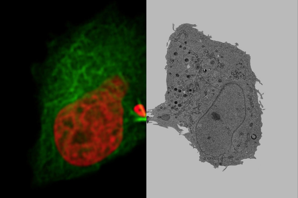
Live Cell CLEM Workflows
Perform experiments on your sample and immediately freeze it to capture the process.
- With EM ICE, precious events can be snap frozen for further investigations under electron microscope.
- Connect live cell imaging with high-pressure freezing to enable live cell CLEM workflows.
Freeze your sample wherever you want
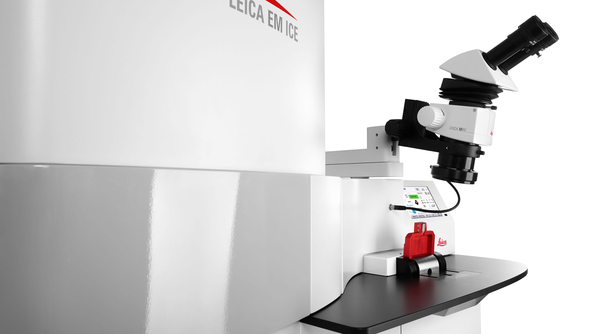
Experience the convenience of a mobile, all-in-one compact instrument.
Easily move EM ICE on wheels and bring it to the sample for fast transfer during live cell CLEM experiments.
Enjoy easy and quick setup, can load the sample with one move, and rapidly start high-pressure freezing.