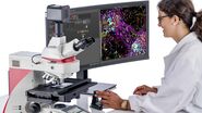
Basis for correct decisions when screening & sorting zebrafish
Do you struggle to see all pigments and fine structural differences in your zebrafish? The identification of the right phenotype is crucial and can be demanding.
See more details – Leica zebrafish screening solutions allow you to recognize fine structures and multiple colors even at low magnification. The acquired results can mean a clearer understanding of the zebrafish nervous system, heart, blood vessels, and pigmentation.
Improve your work experience by
- Easy selection of an optimal contrast;
- Having a homogenous contrast over the entire field of view (FOV);
- Maintaining a steady contrast without re-adjusting over the entire zoom range.
Make a difference when screening & sorting zebrafish with Leica stereo microscopes.

Zebrafish Research Overview articles
Imaging and Analyzing Zebrafish, Medaka, and Xenopus

Using U-Shaped Glass Capillaries for Sample Mounting

Reveal information with different contrast options
Cleary see the non-fluorescent stains in your zebrafish with brightfield stereo microscopy and the Leica transmitted light (TL) bases:
- Evaluate staining and see the realistic colors of your zebrafish;
- Investigate internal structures and visualize fine details with different contrast methods.
Fluorescence screening: Don’t let background noise mask your signals
Detecting xFP fusion proteins can be very challenging. The goal is to keep the expression of fluorescent proteins at low levels for realistic physiology. Signals are, therefore, faint and hard to identify.
In this case, your microscope setup should not be working against you with high levels of autofluorescence.
The Leica solution to this problem:
- A separate excitation channel in fluorescence stereo microscopes, i.e., Triple Beam;
- A specially designed mirror in light bases that helps prevent autofluorescence;
- Adjustable flaps covering the automated light base to minimize autofluorescence during imaging.
The result is a clear fluorescence signal against a noise-free, black background.

Handle your specimen with ease
Being able to easily see the orientation in 3D with Leica stereo microscopes ensures that you see exactly where your tools are. That way, you don’t accidently injure your zebrafish.
With Leica solutions, you can:
- Handle zebrafish specimens easily with a good hand-eye-coordination;
- Set injections and lesions precisely;
- Optimally position your sample for imaging.
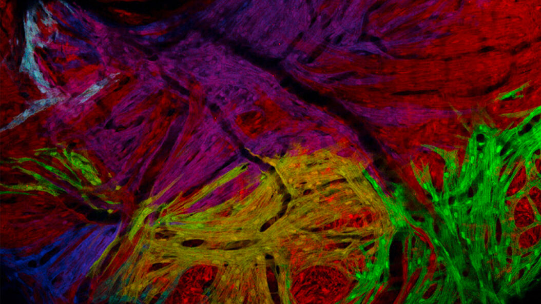
 viewed with Rottermann contrast. Courtesy of Vermot Laboratory, IGBMC Strasbourg, France.")
Efficiently obtain publishable results
Functional imaging of zebrafish specimens is one of the most demanding tasks to perform in the laboratory. Researchers can achieve it by taking advantage of Leica automated stereo fluorescent microscopes.
Work with zebrafish more efficiently thanks to software-based control of:
- Fluorescence excitation light, filters, and shutter
- Transmitted light contrast
- X/Y/Z positioning of specimen
- Microscope camera for image acquisition
- Magnification
- Specimen environment
With a fully automated Leica transmitted light bases, functional imaging of zebrafish is done more easily, as they provide the resolution and contrast required.

FAQs Zebrafish Research
Zebrafish are highly regenerative. They can regrow amputated fins, as well as lesioned brain, retina, spinal cord, heart, and other tissues. Additionally, they are considered to be in vitro, not alive, until 5 days post fertilization (dpf), making them a less controversial alternative for research compared to mammalian model organisms.
The genome of the zebrafish has been fully sequenced. It is easy to genetically manipulate them. Their fecundity is high, fertilization is external, development is rapid, and the embryo is nearly transparent. For these reasons, zebrafish are useful for biomedical and biological research, including studies of cellular processes and diseases.
Humans and zebrafish actually share 70% of the same genes. Many of the genes required to grow the heart, mouth, eyes, kidney, muscle, blood, etc., are conserved between them. Zebrafish offer advantages over mammalian model organisms, like mice and rats, even though they are evolutionarily more similar to humans.
.")