Capturing Developmental Dynamics in 3D
From organoids to embryos: Capturing developmental dynamics in 3D with Viventis Deep light sheet imaging

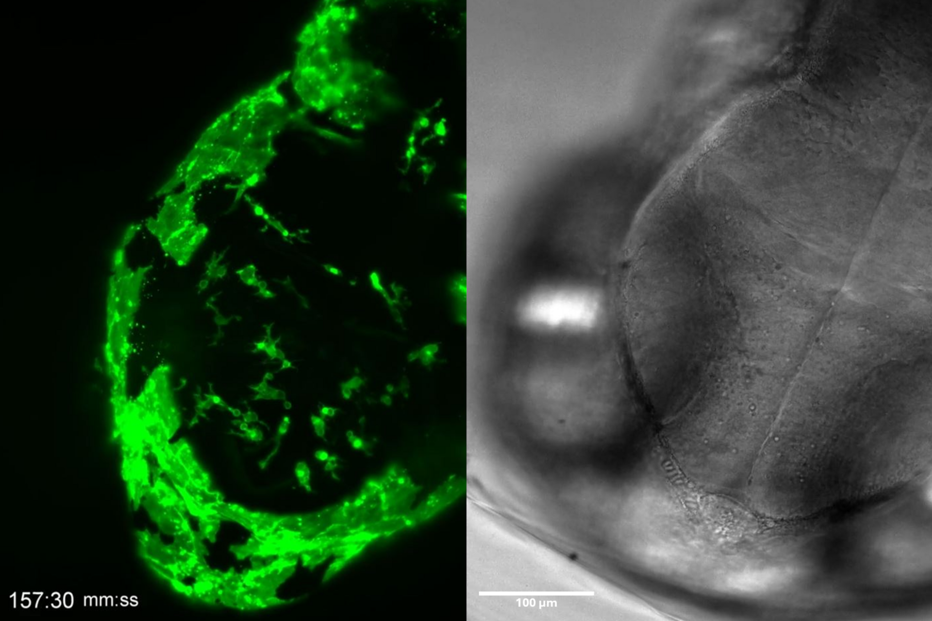
This application note showcases how the Viventis Deep dual-view light sheet microscope was successfully used by researchers for exploring high-resolution, long-term imaging of 3D multicellular models – including organoids, spheroids, and embryos – unlocking new possibilities in developmental biology and disease research.
Relevant applications for light sheet microscopy include:
- 3D models that mimic in vivo conditions, making them valuable for drug screening and toxicology.
- Embryos that are key for studying development and genetic disorders.
- Long-term live imaging, which is essential for observing dynamic processes but faces challenges such as light scattering, phototoxicity, and maintaining physiological conditions.
Light Sheet imaging is the gentlest method to date. Imaging with Viventis Deep means:
- Deeper penetration of the sample with double illumination and dual view detection.
- Easy sample handling and multi-position experiments.
- Easy sample mounting and medium exchange via multi-well, open-top chambers.
- Precise environmental control, supporting long-term imaging of complex 3D samples.
Related Articles
-

History, Developments and Trends of Microscopy in Cancer Research
Cancer is a global disease, with 18 million new cases diagnosed and 10 million cancer-related deaths…
Mar 16, 2026Read article -
 and astrocytes (green) in a cortical spheroid derived from human induced pluripotent stem cells.")
Guide to Live-Cell Imaging
For a wide range of applications in various research fields of life science, live-cell imaging is an…
Jan 12, 2026Read article -
.")
Focus on Long-Term Imaging in 3D with Light Sheet Microscopy
Long-term 3D imaging reveals how complex multicellular systems grow and develop and how cells move…
Oct 06, 2025Read article
