
Larger 3D area in focus during surgery - clear view of anatomical details
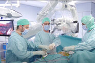
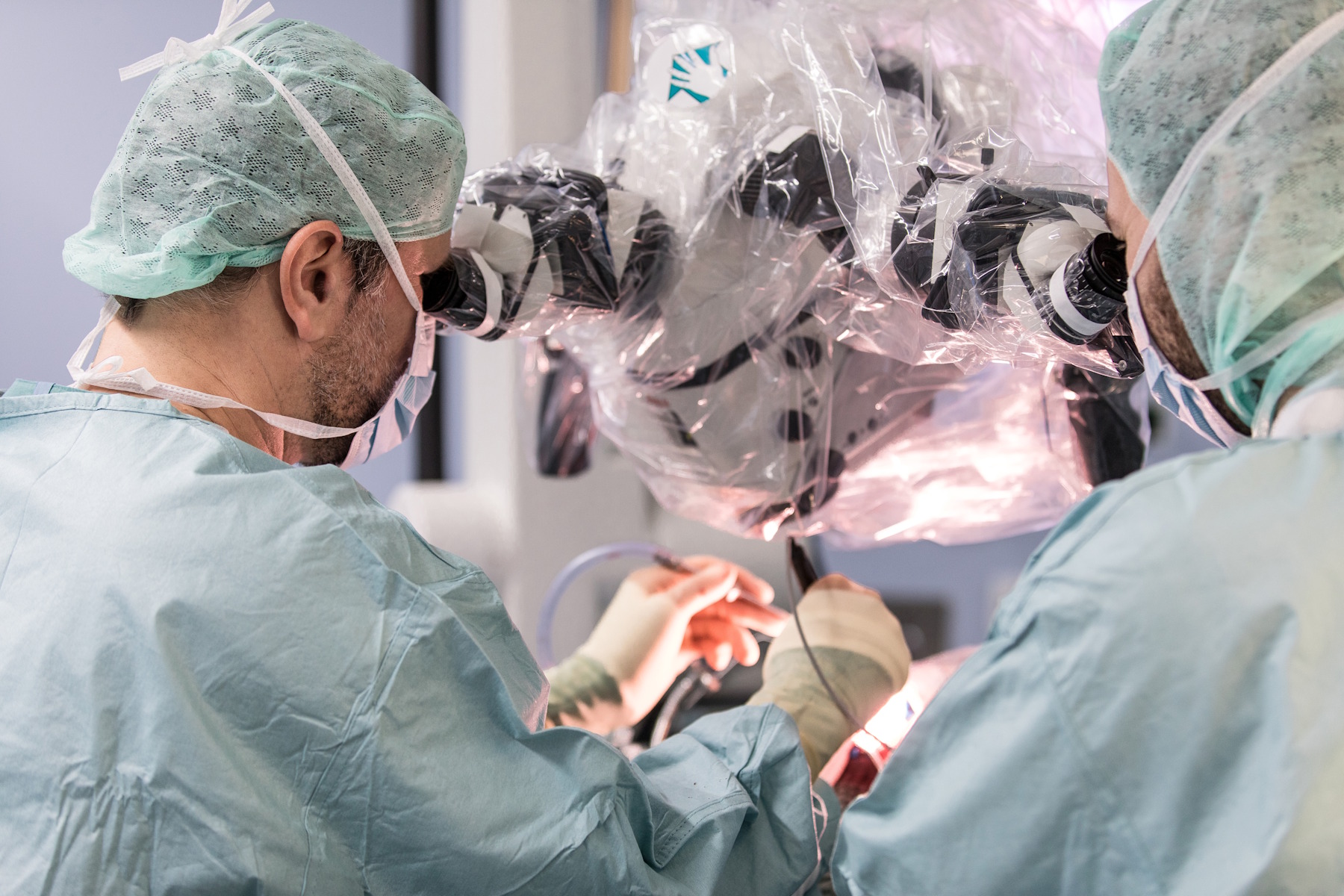
When performing neurosurgery or ophthalmic surgery, having a greater area in focus which eliminates frequent refocusing helps streamline the surgical workflow. In combination with advanced illumination and apochromatic optics, a surgeon can see more anatomical details clearly, especially at the bottom of a deep cavity (refer to video 1 below).
Video 1: Explanation of how surgeons can benefit from a Leica surgical microscope which provides a higher resolution through one eyepiece and a greater depth of field through the other. Each eye observes a different image, and these are combined together by the brain. The result is that the surgeon sees a larger 3D area in focus and a clear view of anatomical details.
Surgical microscopes with more than 30% increased depth of field
For conventional microscope imaging, depth of field is determined by the correlation between numerical aperture and magnification [1-3]. For the best possible visual impression, the parameters of conventional microscopes can be adjusted to produce an optimum balance between depth of field and resolution which, in theory, are inversely correlated. Particularly at low magnifications, the depth of field can be significantly increased by reducing the numerical aperture. However, the smaller the numerical aperture, the lower the lateral resolution. Therefore, it has always been a challenge to find the optimum balance of resolution and depth of field.
However, there is a sophisticated optical approach that resolves the inverse correlation between resolution and depth of field. This stereo-microscope technology, developed by Leica Microsystems, is called FusionOptics [4]. It uses two different-sized apertures in the optical pathways: A large aperture provides high resolution, and a smaller one provides greater depth of field. The human brain combines the two separate images into a single impression that features both high resolution and high depth of field (refer to figure 1). Thus, the depth of field can be increased by over 30%.

Fig. 1: How FusionOptics works: There are two separate beam paths (1) for each eye where one beam path (2) provides greater depth of field and the other path (3) higher resolution. The two images are merged into a single, optimal spatial image (4) by the brain.
During the development of this technology, a study was conducted to investigate the binocular combination of visual signals for humans [4]. The results showed that the brain is capable of using the best information from both eyes in order to compose an optimal spatial image. This fact is true even when each eye provides different information. Today, thousands of Leica surgical microscopes with FusionOptics technology are in use worldwide, including the ARveo 8x and M530 OHX microscope for neurosurgery, Proveo 8 for ophthalmic surgery, and PROvido multidisciplinary surgical microscope.
 case.")