
Key Learnings
- Learn about the different steps of Moyamoya disease surgical treatment and bypass surgery: pre-operative assessment, intra-operative course and post-surgery assessment
- Gain insights on the impact of GLOW800 Augmented Reality fluorescence in Moyamoya disease treatment and how it improves confidence
- Discover clinical images and exclusive videos of Moyamoya disease surgical management
Moyamoya disease patient initial presentation & pre-operative assessment
The case study looks at a 35-year-old female patient with Moyamoya disease who presented with worsening neurological signs and symptoms. The decision was made to perform bypass surgery.

Before the bypass surgery, a pre-operative assessment was performed including catheter angiography and digital subtraction angiography (DSA). This allowed to better understand the occlusions and stenosis.
Moyamoya disease surgical approach: What is the treatment for Moyamoya disease?
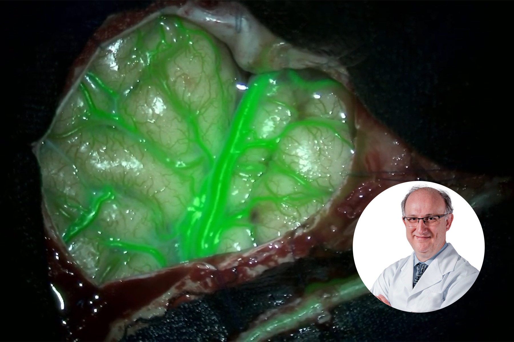
As a first step, the surgeon dissected the superficial artery. The use of GLOW800 Augmented Reality fluorescence enables the assessment of the blood flow through the artery. It also allows to check for potential hemorrhaging and thrombosis.

A pterional craniotomy was performed, followed by the subarachnoid dissection through the Sylvian fissure.
Prior to the anastomosis, GLOW800 was used to assess the cerebral blood flow through the feeding arteries and the M4 branch. The anastomotic mouth of the temporal artery was performed for the bypass.

After completing the bypass, GLOW800 was instrumental to assess the patency of the arteries and ensure the absence of hemorrhages and thrombosis. The postoperative evaluation showed no signs of postoperative complications or new neurological deficits.
Impact of GLOW800 Augmented Reality Fluorescence in Moyamoya disease surgical treatment

The use of GLOW800 Augmented Reality fluorescence provides one augmented real-time view during vascular neurosurgery. Neurosurgeons do not need to recall and try to reconcile the black and white blood flow video with the natural anatomical view. The crisp delineation, full depth perception and absence of dark peripheries supports precise manipulation of vessels.
Benefits of GLOW800 Augmented Reality
With GLOW800 Augmented Reality, it is possible to:
- Perform an accurate assessment of blood flow during bypass surgery through the feeding and receiving vessels, aiding in their selection
- Identify the presence of thrombosis or stenosis of vessels before anastomosis and do an intraoperative assessment of the patency of anastomosis
- Intraoperatively assess blood hemorrhage through anastomosed blood vessels, including kinking and partial obstruction.
As such, GLOW800 supports Moyamoya disease treatment and outcomes and is a fundamental tool for the future of practice and training in neurosurgery. Want to learn more? Register below to download the full case study and access the clinical videos.
Note: The statements of the healthcare professional in this case study reflect only his opinion and personal experience. His statements do not necessarily reflect the opinion of any institution with whom he is affiliated. Please check with your local Leica Microsystems representative for product registration status in your region.
Related Articles
-

Enhancing Neurosurgery Teaching
Learn about the Serious Game in Intraoperative Neurosurgery and how it supports neurosurgical…
Oct 12, 2023Read article -

Launching a Neurosurgical Department with Limited Resources
Learn about Dr. Claire Karekezi’s journey and experience launching a neurosurgical department within…
Sep 11, 2023Read article -

Use of AR Fluorescence in Neurovascular Surgery
Learn about the use of GLOW800 Augmented Reality in neurovascular surgery through clinical cases and…
Aug 02, 2023Read article
