
Science Lab
Science Lab
The knowledge portal of Leica Microsystems offers scientific research and teaching material on the subjects of microscopy. The content is designed to support beginners, experienced practitioners and scientists alike in their everyday work and experiments. Explore interactive tutorials and application notes, discover the basics of microscopy as well as high-end technologies – become part of the Science Lab community and share your expertise!
Filter articles
Tags
Products
Loading...

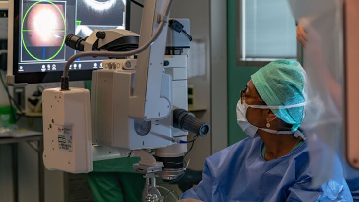
Buying an Ophthalmic Microscope? Gain Peers Insights from Dr. Dhami
In this article, learn how Dr. Abhinav Dhami, an ophthalmic surgery consultant in North India, enhances his surgical precision using the M822 ophthalmic microscope from Leica Microsystems and the key…
Loading...
 set-up.")
Augmented Reality: Transforming Neurosurgical Procedures
In this ebook, you will explore the exciting advances that Augmented Reality (AR) brings to the field of neurosurgery. This comprehensive guide, including explanatory videos, addresses key questions…
Loading...

Tympanoplasty Surgery: Optimal Approaches and Tools
Discover tympanoplasty surgery case studies illustrating the standard approaches: post-auricular, endaural and transcanal. Gain insights from Dr. Flanagan on selecting the adequate surgical approach…
Loading...
RPE65 Gene Therapy Subretinal Injection: Benefits of Intraoperative OCT
Discover how RPE65 gene therapy subretinal injection procedures in patients with Leber congenital amaurosis is supported by intraoperative Optical Coherence Tomography.
Loading...

Posterior Segment Surgery: Benefits of Utilizing Intraoperative OCT
Learn about the value of intraoperative optical coherence tomography in posterior segment surgery to precisely locate, evaluate and manage pathologies.
Loading...

Intraoperative OCT-Assisted Corneal Transplant Procedures
Learn about the use of intraoperative optical coherence tomography in corneal transplantation and how it facilitates the adaptation of the donor cornea.
Loading...

Enhancing Neurosurgery Teaching
Learn about the Serious Game in Intraoperative Neurosurgery and how it supports neurosurgical teaching and the acquisition of decision-making skills.
Loading...

How Intraoperative OCT Helps Gain Greater Insight in Glaucoma Surgery
Learn about the use of intraoperative Optical Coherence Tomography in glaucoma surgery and how it helps see subsurface tissue details.
Loading...

Ophthalmology: Visualization in Complex Cataract Surgery
Learn about the use of intraoperative Optical Coherence Tomography in cataract surgery and how it supports both standard and complex cataract surgery cases.