
Science Lab
Science Lab
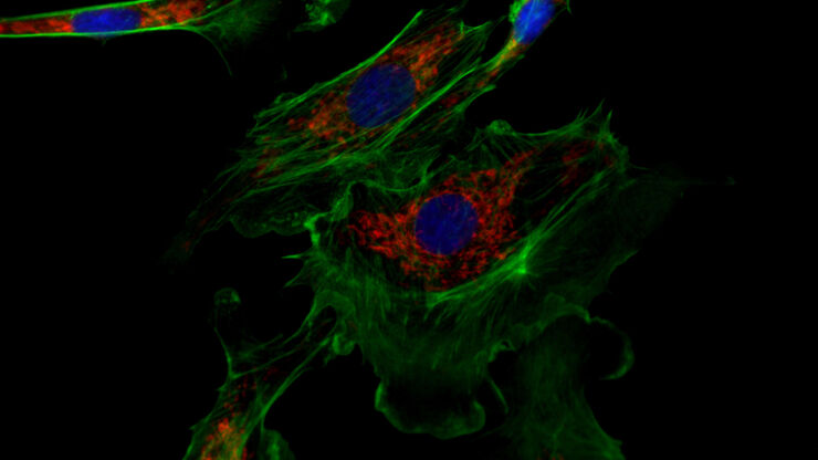
The knowledge portal of Leica Microsystems offers scientific research and teaching material on the subjects of microscopy. The content is designed to support beginners, experienced practitioners and scientists alike in their everyday work and experiments. Explore interactive tutorials and application notes, discover the basics of microscopy as well as high-end technologies – become part of the Science Lab community and share your expertise!
Filter articles
Tags
Products
Loading...

Empowering Spatial Biology with Open Multiplexing and Cell DIVE
Spatial biology and multiplexed imaging workflows have become important in immuno-oncology research. Many researchers struggle with study efficiency, even with effective tools and protocols. Here, we…
Loading...

How did Laser Microdissection enable Pioneering Neuroscience Research?
Dr. Marta Paterlini, a Senior Scientist at the Karolinska Institute, shares her experience of using laser microdissection (LMD) in groundbreaking research into adult human neurogenesis and offers…
Loading...

How do Cells Talk to Each Other During Neurodevelopment?
Professor Silvia Capello presents her group’s research on cellular crosstalk in neurodevelopmental disorders, using models such as cerebral organoids and assembloids.
Loading...

Overcoming Observational Challenges in Organoid 3D Cell Culture
Learn how to overcome challenges in observing organoid growth. Read this article and discover new solutions for real-time monitoring which do not disturb the 3D structure of the organoids over time.
Loading...

Technical Terms for Digital Microscope Cameras and Image Analysis
Learn more about the basic principles behind digital microscope camera technologies, how digital cameras work, and take advantage of a reference list of technical terms from this article.
Loading...

The Shape of the Brain: Spatial Biology of Alzheimer’s Disease
Uncover cell identity and brain structure in Alzheimer's disease with Cell DIVE multiplexed imaging, demonstrating how spatial biology can lead to advances in therapy development for…
Loading...
Studying Virus Replication with Fluorescence Microscopy
The results from research on SARS-CoV-2 virus replication kinetics, adaption capabilities, and cytopathology in Vero E6 cells, done with the help of fluorescence microscopy, are described in this…
Loading...
")
Introduction to Fluorescent Proteins
Overview of fluorescent proteins (FPs) from, red (RFP) to green (GFP) and blue (BFP), with a table showing their relevant spectral characteristics.
Loading...

ISO 9022 Standard Part 11 - Testing Microscopes with Severe Conditions
This article describes a test to determine the robustness of Leica microscopes to mold and fungus growth. The test follows the specifications of the ISO 9022 part 11 standard for optical instruments.