
Science Lab
Science Lab
The knowledge portal of Leica Microsystems offers scientific research and teaching material on the subjects of microscopy. The content is designed to support beginners, experienced practitioners and scientists alike in their everyday work and experiments. Explore interactive tutorials and application notes, discover the basics of microscopy as well as high-end technologies – become part of the Science Lab community and share your expertise!
Filter articles
Tags
Products
Loading...
Confocal Imaging of Immune Cells in Tissue Samples
In this webinar, you will discover how to perform 10-color acquisition using a confocal microscope. The challenges of imaged-based approaches to identify skin immune cells. A new pipeline to assess…
Loading...
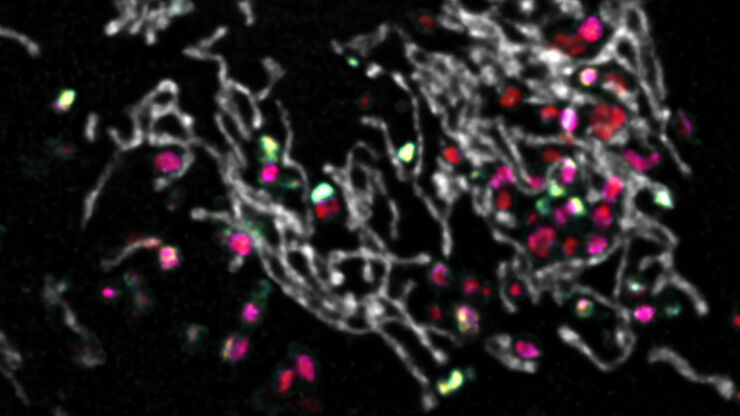
Insights into Vesicle Trafficking
STELLARIS provides integral access to complementary layers of information for dynamic, structural, and mechanistic insights into vesicle trafficking.
Loading...
Visualizing Protein-Protein Interactions by Non-Fitting and Easy FRET-FLIM Approaches
The Webinar with Dr. Sergi Padilla-Parra is about visualizing protein-protein interaction. He gives insight into non-fitting and easy FRET-FLIM approaches.
Loading...

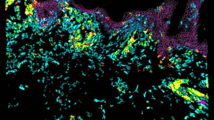
Multiplexing through Spectral Separation of 11 Colors
Fluorescence microscopy is a fundamental tool for life science research that has evolved and matured together with the development of multicolor labeling strategies in cells tissues and model…
Loading...
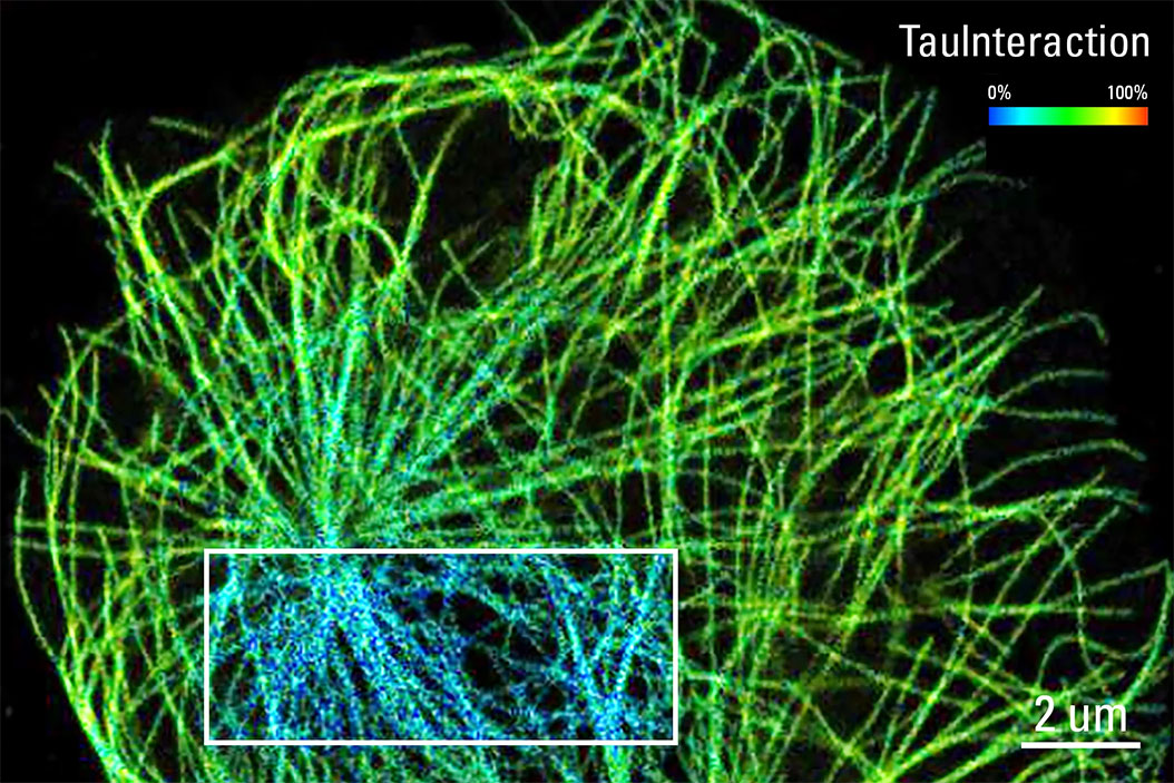
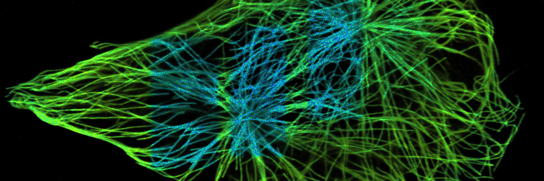
TauInteraction – Studying Molecular Interactions with TauSense
Fluorescence microscopy constitutes one of the pillars in life sciences and is a tool commonly used to unveil cellular structure and function. A key advantage of fluorescence microscopy resides in the…
Loading...

Find Relevant Specimen Details from Overviews
Switch from searching image by image to seeing the full overview of samples quickly and identifying the important specimen details instantly with confocal microscopy. Use that knowledge to set up…
Loading...

Benefits of TauContrast to Image Complex Samples
In this interview, Dr. Timo Zimmermann talks about his experience with the application of TauSense tools and their potential for the investigation of demanding samples such as thick samples or…
Loading...
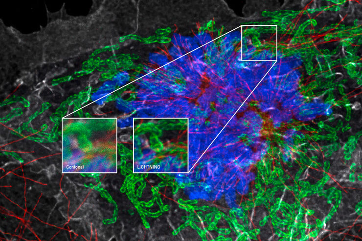
Obtain Maximum Information from your Specimen with LIGHTNING
LIGHTNING is an adaptive process for extraction of information that reveals fine structures and details, otherwise simply not visible, fully automatically. Unlike traditional technologies, that use a…
Loading...
Explore Innovative Techniques to Separate Fluorophores with Overlapping Spectra
In this article we explore several strategies you can take to improve the separation of fluorophores and increase the number of fluorescent probes you can distinguish in your sample.