
Science Lab
Science Lab
The knowledge portal of Leica Microsystems offers scientific research and teaching material on the subjects of microscopy. The content is designed to support beginners, experienced practitioners and scientists alike in their everyday work and experiments. Explore interactive tutorials and application notes, discover the basics of microscopy as well as high-end technologies – become part of the Science Lab community and share your expertise!
Filter articles
Tags
Products
Loading...
Fluorescence Lifetime-based Imaging Gallery
Confocal microscopy relies on the effective excitation of fluorescence probes and the efficient collection of photons emitted from the fluorescence process. One aspect of fluorescence is the emission…
Loading...

Physiology Image Gallery
Physiology is about the processes and functions within a living organism. Research in physiology focuses on the activities and functions of an organism’s organs, tissues, or cells, including the…
Loading...

Live Cell Imaging Gallery
Live cell microscopy techniques are fundamental to get a better understanding of cellular and molecular function. Today, widefield microscopy is the most common technique used to visualize cell…
Loading...
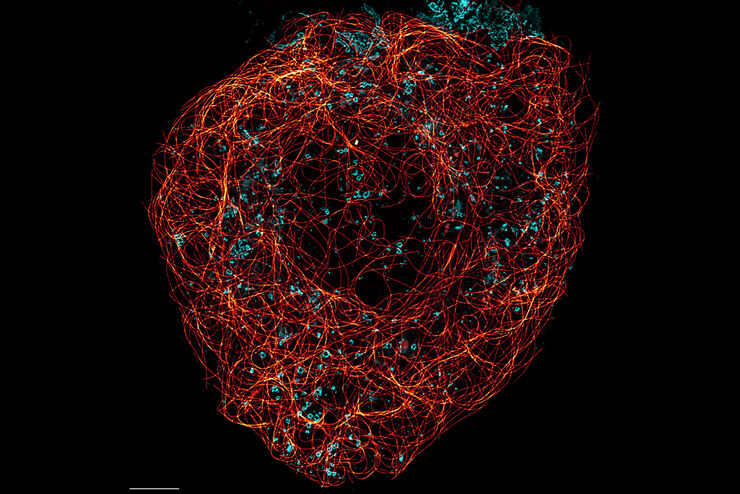
Super-Resolution Microscopy Image Gallery
Due to the diffraction limit of light, traditional confocal microscopy cannot resolve structures below ~240 nm. Super-resolution microscopy techniques, such as STED, PALM or STORM or some…
Loading...
Tissue Image Gallery
Visual analysis of animal and human tissues is critical to understand complex diseases such as cancer or neurodegeneration. From basic immunohistochemistry to intravital imaging, confocal microscopy…
Loading...
 and astrocytes (green) in a cortical spheroid derived from human induced pluripotent stem cells.")
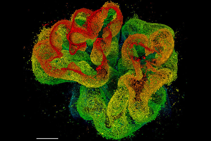
Neuroscience Images
Neuroscience commonly uses microscopy to study the nervous system’s function and understand neurodegenerative diseases.
Loading...
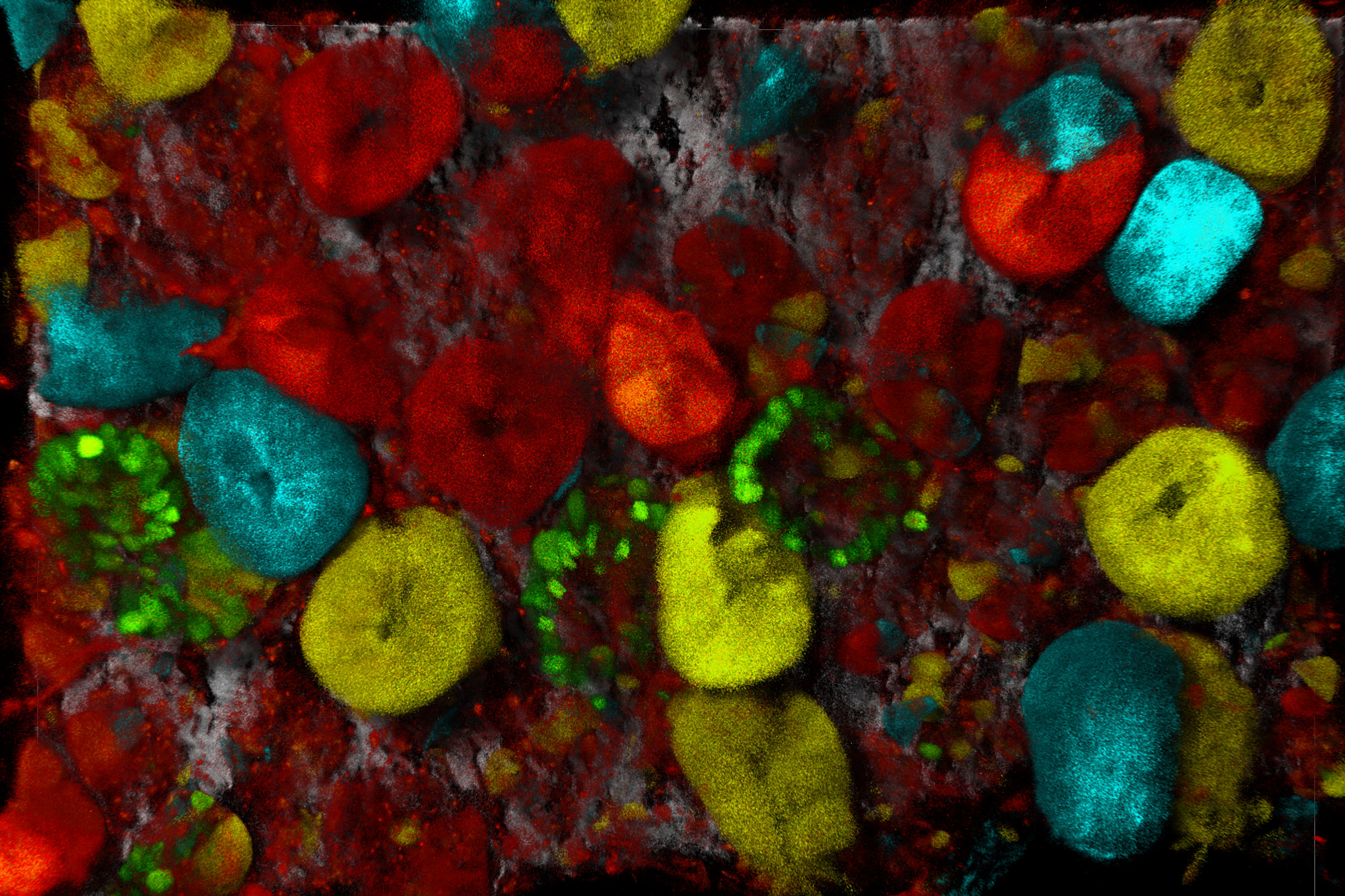
Multicolor Image Gallery
Fluorescence multicolor microscopy, which is one aspect of multiplex imaging, allows for the observation and analysis of multiple elements within the same sample – each tagged with a different…
Loading...
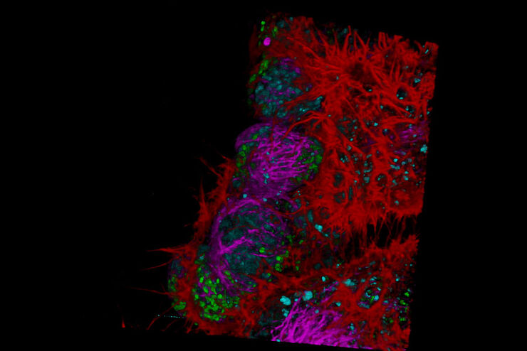
Cancer Research Image Gallery
Fluorescence microscopy allows the study of changes occurring in tissue and cells during cancer development and progression. Techniques such as live cell imaging are critical to understand cancer…
Loading...
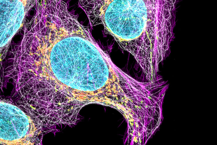
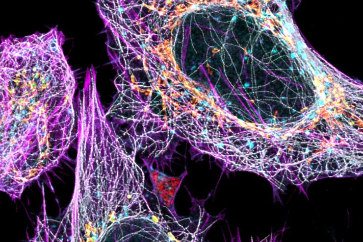
Cell Biology Image Gallery
Cell biology studies the structure, function and behavior of cells, including cell metabolism, cell cycle, and cell signaling. Fluorescence microscopes are an integral part of a cell biologist…
