
Science Lab
Science Lab
The knowledge portal of Leica Microsystems offers scientific research and teaching material on the subjects of microscopy. The content is designed to support beginners, experienced practitioners and scientists alike in their everyday work and experiments. Explore interactive tutorials and application notes, discover the basics of microscopy as well as high-end technologies – become part of the Science Lab community and share your expertise!
Filter articles
Tags
Products
Loading...

Understanding Clearly the Magnification of Microscopy
To help users better understand the magnification of microscopy and how to determine the useful range of magnification values for digital microscopes, this article provides helpful guidelines.
Loading...
 of an image of two points where the distance between them corresponds to the Rayleigh criterion.")
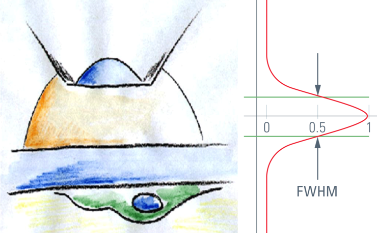
Microscope Resolution: Concepts, Factors and Calculation
This article explains in simple terms microscope resolution concepts, like the Airy disc, Abbe diffraction limit, Rayleigh criterion, and full width half max (FWHM). It also discusses the history.
Loading...

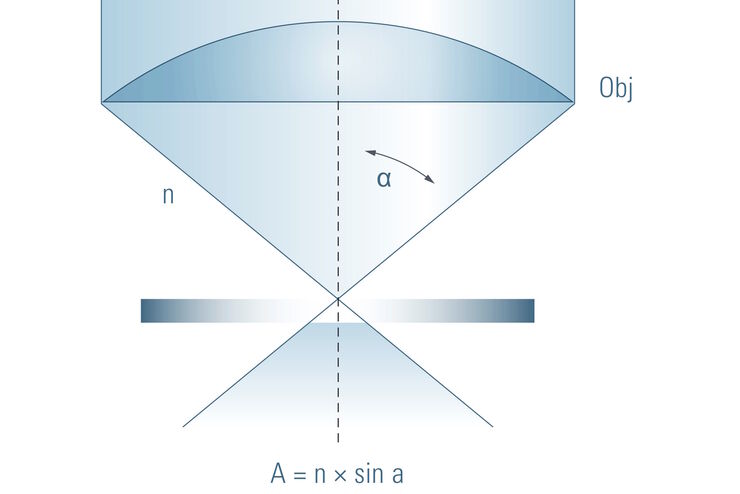
Collecting Light: The Importance of Numerical Aperture in Microscopy
Numerical aperture (abbreviated as ‘NA’) is an important consideration when trying to distinguish detail in a specimen viewed down the microscope. NA is a number without units and is related to the…
Loading...
")
A Brief History of Light Microscopy
The history of microscopy begins in the Middle Ages. As far back as the 11th century, plano-convex lenses made of polished beryl were used in the Arab world as reading stones to magnify manuscripts.…
Loading...

Optical Microscopes – Some Basics
The optical microscope has been a standard tool in life science as well as material science for more than one and a half centuries now. To use this tool economically and effectively, it helps a lot to…
Loading...
Confocal Optical Section Thickness
Confocal microscopes are employed to optically slice comparably thick samples.
Loading...

Depth of Field in Microscopy
In microscopy, depth of field is often seen as an empirical parameter. In practice it is determined by the correlation between numerical aperture, resolution and magnification. For the best possible…
Loading...
Beware of "Empty" Magnification
This article explains how to avoid the phenomen of "empty magnification" in microscopy.