
Science Lab
Science Lab
The knowledge portal of Leica Microsystems offers scientific research and teaching material on the subjects of microscopy. The content is designed to support beginners, experienced practitioners and scientists alike in their everyday work and experiments. Explore interactive tutorials and application notes, discover the basics of microscopy as well as high-end technologies – become part of the Science Lab community and share your expertise!
Filter articles
Tags
Products
Loading...

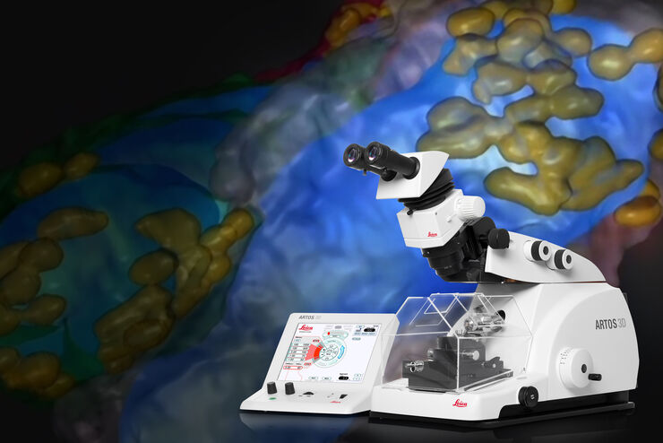
Automatic Alignment of Sample and Knife for High Sectioning Quality
Automatic alignment of sample and knife on the ultramicrotome UC Enuity, enabling even untrained users to create ultrathin sections with reduced risk of losing precious sections.
Loading...

High Quality Sectioning in Ultramicrotomy
Discover the significance of achieving high-quality uniform sections with ultramicrotomy for precise imaging in electron microscopy.
Loading...

Streamline your EM Sample Preparation Workflow for Biological Applications
Master EM sample preparation, including ultramicrotomy, for life sciences in this expert eBook!
Loading...
 stained with osmium tetroxide (OsO4), sectioned with a DIATOME diamond knife at room temperature, and then imaged with HAADF TEM.")
Ultramicrotomy Techniques for Materials Sectioning
Learn about ultramicrotomy for materials sectioning when investigating polymers and brittle materials with transmission (TEM) or scanning electron microscopy (SEM) or atomic force microscopy.
Loading...

Introduction to Ultramicrotomy
When studying samples, to visualize their fine structure with nanometer scale resolution, most often electron microscopy is used. There are 2 types: scanning electron microscopy (SEM) which images the…
Loading...
Array Tomography for SEM 3D Reconstruction
Array tomography (AT) is a 3D image reconstruction technique for high resolution, quantitative analysis of biological structures. For optimal results, ultrathin and ordered sections are an absolute…
Loading...
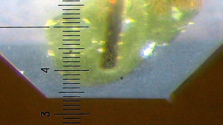
Brief Introduction to Specimen Trimming
Before ultrathin sectioning a sample with an ultramicrotome it has to be pre-prepared. For this pre-preparation, special attention must be paid to the sample size (size of the section), location of…