
Science Lab
Science Lab
The knowledge portal of Leica Microsystems offers scientific research and teaching material on the subjects of microscopy. The content is designed to support beginners, experienced practitioners and scientists alike in their everyday work and experiments. Explore interactive tutorials and application notes, discover the basics of microscopy as well as high-end technologies – become part of the Science Lab community and share your expertise!
Filter articles
Tags
Products
Loading...
 and oblique (right) brightfield illumination using a Leica compound microscope. The defect on the wafer surface is clearly more visible with oblique illumination.")
Rapid Semiconductor Inspection with Microscope Contrast Methods
Semiconductor inspection for QC of materials like wafers can be challenging. Microscope solutions that offer several contrast methods enable fast and reliable defect detection and efficient workflows.
Loading...
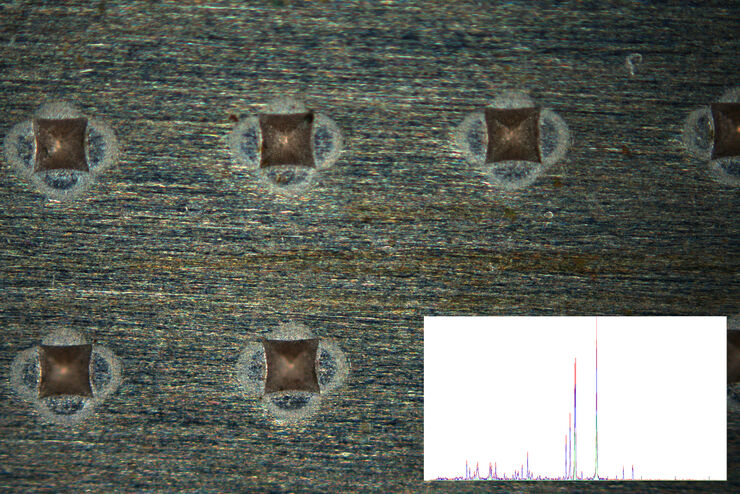
Fast Visual and Chemical Analysis of Contamination and Underlying Layers
Visual and chemical analysis of contamination on materials with a 2-methods-in-1 solution leading to an efficient, more complete analysis workflow is described in this report. A 2-in-1 solution,…
Loading...
Metallography – an Introduction
This article gives an overview of metallography and metallic alloy characterization. Different microscopy techniques are used to study the alloy microstructure, i.e., microscale structure of grains,…
Loading...

How to Adapt Grain Size Analysis of Metallic Alloys to Your Needs
Metallic alloys, such as steel and aluminum, have an important role in a variety of industries, including automotive and transportation. In this report, the importance of grain size analysis for alloy…
Loading...

Imaging and Analyzing Zebrafish, Medaka, and Xenopus
Discover how to image and analyze zebrafish, medaka, and Xenopus frog model organisms efficiently with a microscope for developmental biology applications from this article.
Loading...

Studying Caenorhabditis elegans (C. elegans)
Find out how you can image and study C. elegans roundworm model organisms efficiently with a microscope for developmental biology applications from this article.
Loading...
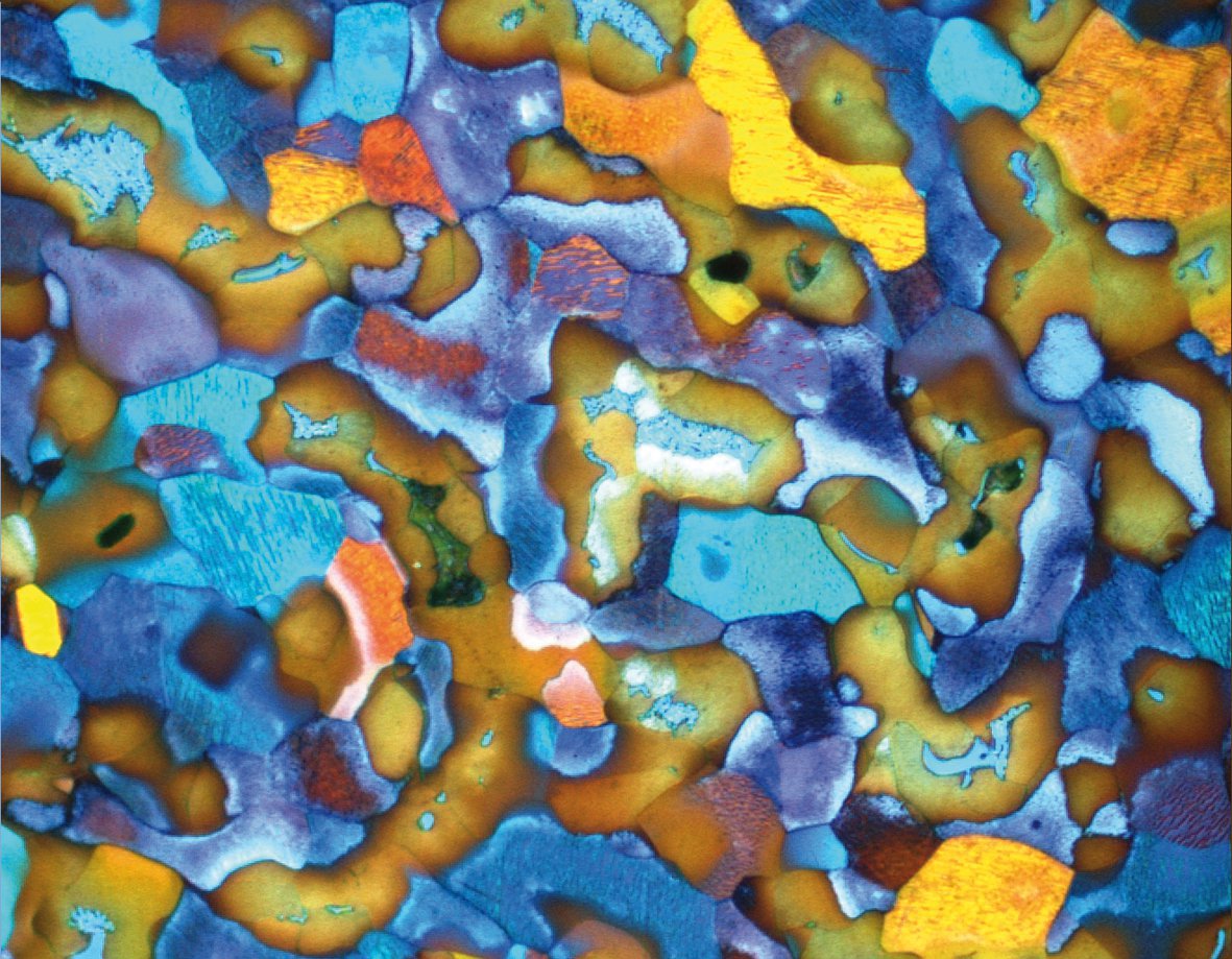
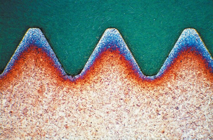
Metallography with Color and Contrast
The examination of microstructure morphology plays a decisive role in materials science and failure analysis. There are many possibilities of visualizing the real structures of materials in the light…
Loading...

Optical Contrast Methods
Optical contrast methods give the potential to easily examine living and colorless specimens. Different microscopic techniques aim to change phase shifts caused by the interaction of light with the…