
Science Lab
Science Lab
The knowledge portal of Leica Microsystems offers scientific research and teaching material on the subjects of microscopy. The content is designed to support beginners, experienced practitioners and scientists alike in their everyday work and experiments. Explore interactive tutorials and application notes, discover the basics of microscopy as well as high-end technologies – become part of the Science Lab community and share your expertise!
Filter articles
Tags
Products
Loading...

Transforming Multiplexed 2D Data into Spatial Insights Guided by AI
Aivia 13 handles large 2D images and enables researchers to obtain deep insights into microenvironment surrounding their phenotypes with millions of detected objects and automatic clustering up to 30…
Loading...

AI Microscopy Image Analysis – An Introduction
Artificial intelligence-guided microscopy image analysis and visualization is a powerful tool for data-driven scientific discovery. AI can help researchers tackle challenging imaging applications,…
Loading...
Using Machine Learning in Microscopy Image Analysis
Recent exciting advances in microscopy technologies have led to exponential growth in quality and quantity of image data captured in biomedical research. However, analyzing large and increasingly…
Loading...
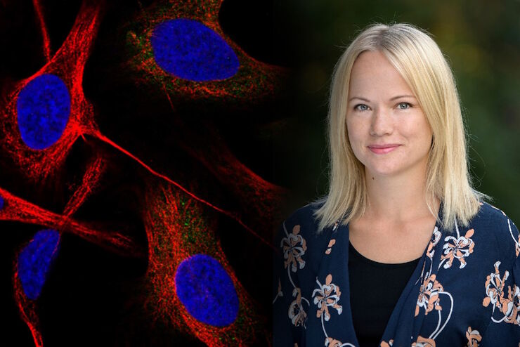
Applying AI and Machine Learning in Microscopy and Image Analysis
Prof. Emma Lundberg is a professor in cell biology proteomics at KTH Royal Institute of Technology, Sweden. She is also the director of the Cell Atlas, an integral part of the Swedish-based Human…
Loading...
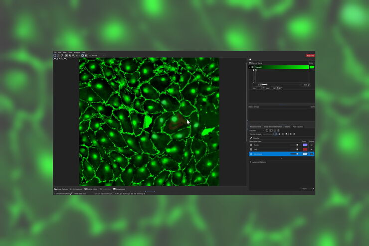
The AI-Powered Pixel Classifier
Achieving reproducible results manually requires expertise and is tedious work. But now there is a way to overcome these challenges by speeding up this analysis to extract the real value of the image…
Loading...
 enhanced with Aivia’s Pixel Classifier (right)")
Simplifying the Cancer Biology Image Analysis Workflow
As cancer biology data sets grow, so do the challenges in microscopy image analysis. Aivia experts cover how to overcome these challenges with AI.
Loading...

Observing Complex Cellular Interactions at Multiple Scales
Learn how to observe challenging cellular interactions with easy to deploy object detection and relationship measurements.
Loading...
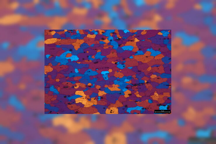
Microstructural Characterization including Compositional Analysis
Leica Microsystems' versatile upright compound microscope, DM6 M, fitted with Laser-Induced Breakdown Spectroscopy module will let you not only analyze metallographically polished samples and conduct…
Loading...

Physiology Image Gallery
Physiology is about the processes and functions within a living organism. Research in physiology focuses on the activities and functions of an organism’s organs, tissues, or cells, including the…