
Science Lab
Science Lab
The knowledge portal of Leica Microsystems offers scientific research and teaching material on the subjects of microscopy. The content is designed to support beginners, experienced practitioners and scientists alike in their everyday work and experiments. Explore interactive tutorials and application notes, discover the basics of microscopy as well as high-end technologies – become part of the Science Lab community and share your expertise!
Filter articles
Tags
Products
Loading...
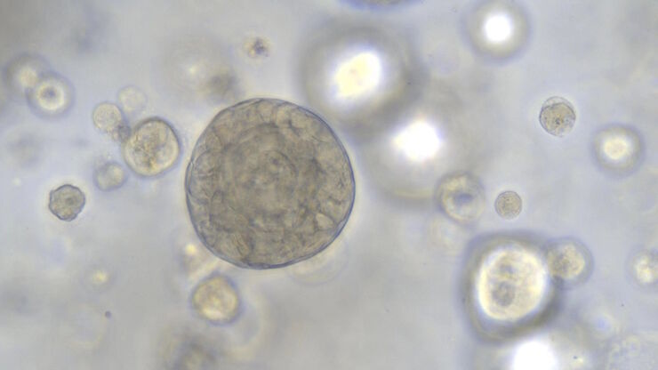
Overcoming Observational Challenges in Organoid 3D Cell Culture
Learn how to overcome challenges in observing organoid growth. Read this article and discover new solutions for real-time monitoring which do not disturb the 3D structure of the organoids over time.
Loading...
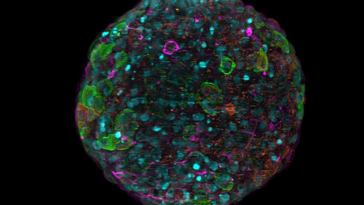
Notable AI-based Solutions for Phenotypic Drug Screening
Learn about notable optical microscope solutions for phenotypic drug screening using 3D-cell culture, both planning and execution, from this free, on-demand webinar.
Loading...
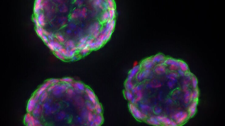
How to Get Deeper Insights into your Organoid and Spheroid Models
In this eBook, learn about key considerations for imaging 3D cultures, such as organoids and spheroids, and discover microscopy solutions to shed new insights into dynamic processes in 3D real-time
Loading...
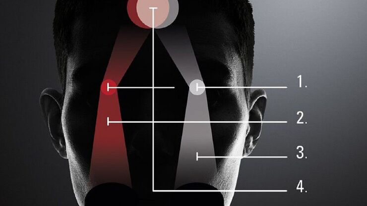
What is the FusionOptics Technology?
Leica stereo microscopes with FusionOptics provide optimal 3D perception. The brain merges two images, one with large depth of field and the other with high resolution, into one 3D image.
Loading...
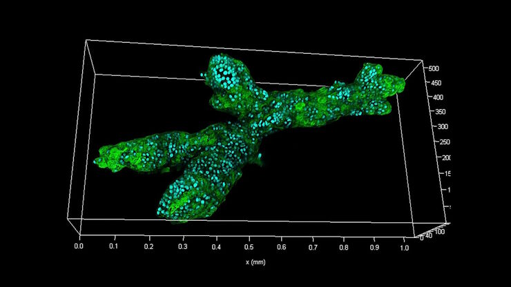
Examining Developmental Processes In Cancer Organoids
Interview: Prof. Bausch and Dr. Pastucha, Technical University of Munich, discuss using microscopy to study development of organoids, stem cells, and other relevant disease models for biomedical…
Loading...
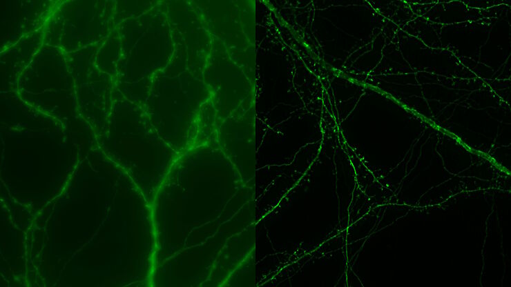
How Microscopy Helps the Study of Mechanoceptive and Synaptic Pathways
In this podcast, Dr Langenhan explains how microscopy helps his team to study mechanoceptive and synaptic pathways, their challenges, and how they overcome them.
Loading...
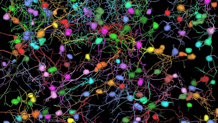
Unlocking Insights in Complex and Dense Neuron Images Guided by AI
The latest advancement in Aivia AI image analysis software provides improved soma detection, additional flexibility in neuron tracing, 3D relational measurement including Sholl analysis and more.
Loading...
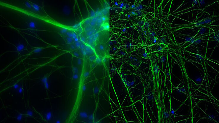
What are the Challenges in Neuroscience Microscopy?
eBook outlining the visualization of the nervous system using different types of microscopy techniques and methods to address questions in neuroscience.
Loading...
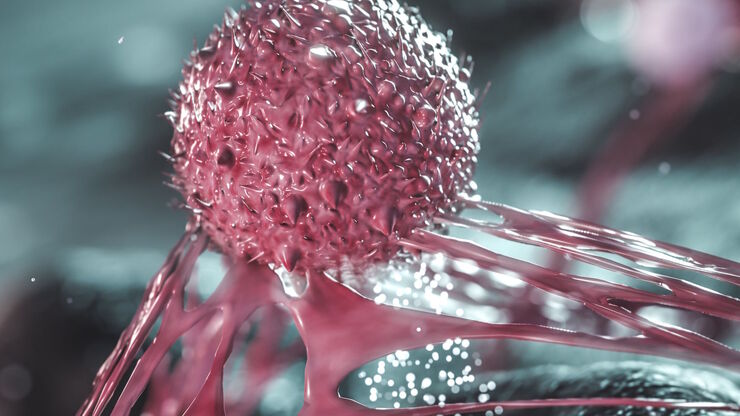
The Role of Iron Metabolism in Cancer Progression
Iron metabolism plays a role in cancer development and progression, and modulates the immune response. Understanding how iron influences cancer and the immune system can aid the development of new…