
Science Lab
Science Lab
The knowledge portal of Leica Microsystems offers scientific research and teaching material on the subjects of microscopy. The content is designed to support beginners, experienced practitioners and scientists alike in their everyday work and experiments. Explore interactive tutorials and application notes, discover the basics of microscopy as well as high-end technologies – become part of the Science Lab community and share your expertise!
Filter articles
Tags
Products
Loading...

Streamline your EM Sample Preparation Workflow for Biological Applications
Master EM sample preparation, including ultramicrotomy, for life sciences in this expert eBook!
Loading...

New Imaging Tools for Cryo-Light Microscopy
New cryo-light microscopy techniques like LIGHTNING and TauSense fluorescence lifetime-based tools reveal structures for cryo-electron microscopy.
Loading...

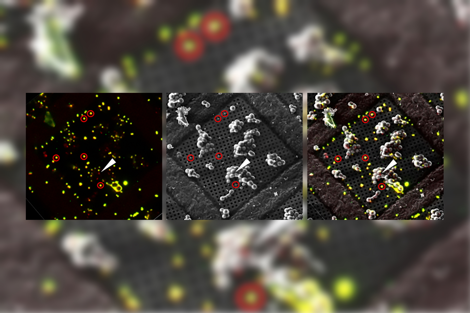
How to Target Fluorescent Structures in 3D for Cryo-FIB Milling
This article describes the major steps of the cryo-electron tomography workflow including super-resolution cryo-confocal microscopy. We describe how subcellular structures can be precisely located in…
Loading...

Precise 3D Targeting for EM Imaging - Access What Matters
Find out how the seamless cryo-electron tomography workflow Coral Cryo uses confocal super resolution to target your structure of interest more precisely.
Loading...
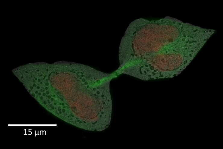
How to Successfully Perform Live-cell CLEM
The Leica Nano workflow provides a streamlined live-cell CLEM solution for getting insight bout structural changes of cellular components over time. Besides the technical handling described in the…
Loading...
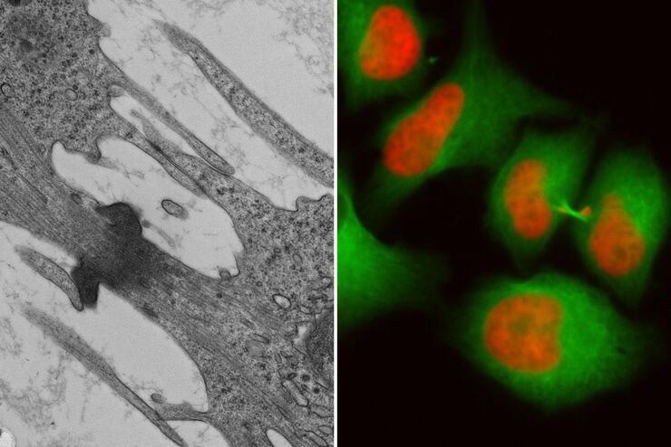
How to Successfully Implement Coral Life
The live-cell CLEM workflow allows you to capture dynamic information related to a relevant biological process as it happens and put these observations into their ultrastructural context. The Leica…
Loading...
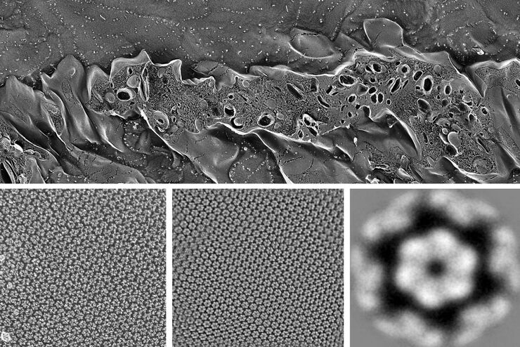
Advancing Cellular Ultrastructure Research
Freeze-fracture and freeze-etching are useful tools for studying flexible membrane-associated structures such as tight junctions or the enteric glycocalyx. Freeze-fracture and etching are two…
Loading...
The Cryo-CLEM Journey
This article describes the Cryo-CLEM technology and the benefits it can provide for scientists. Additionally, some scientific publications are highlighted.
Recent developments in cryo electron…
Loading...

How to Keep Your Samples Under Physiological Conditions
The Coral Life workflow combines dynamic data with the best possible sample fixation by high pressure freezing. However, good sample preservation won’t help if your cells are stressed by temperature…