
Science Lab
Science Lab
The knowledge portal of Leica Microsystems offers scientific research and teaching material on the subjects of microscopy. The content is designed to support beginners, experienced practitioners and scientists alike in their everyday work and experiments. Explore interactive tutorials and application notes, discover the basics of microscopy as well as high-end technologies – become part of the Science Lab community and share your expertise!
Filter articles
Tags
Products
Loading...

Transforming Multiplexed 2D Data into Spatial Insights Guided by AI
Aivia 13 handles large 2D images and enables researchers to obtain deep insights into microenvironment surrounding their phenotypes with millions of detected objects and automatic clustering up to 30…
Loading...

Accelerating Neuron Image Analysis with Automation
The ability to examine complex neural processes relies on the accurate reconstruction of neuronal networks at scale. Most data extraction methods in neuroscience research are time-consuming and…
Loading...
 in 3D")
How Artificial Intelligence Enhances Confocal Imaging
In this article, we show how artificial intelligence (AI) can enhance your imaging experiments. Namely, how Dynamic Signal Enhancement powered by Aivia improves image quality while capturing the…
Loading...

An Introduction to Computational Clearing
Many software packages include background subtraction algorithms to enhance the contrast of features in the image by reducing background noise. The most common methods used to remove background noise…
Loading...
 images")
How To Create EDOF (Extended Depth of Focus) Images
Watch this video to see how you can rapidly record sharp optical microscope images of samples with a large height variation. This is done with the optional Extended Depth of Focus (EDOF) function of…
Loading...
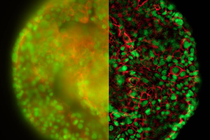
Real Time Images of 3D Specimens with Sharp Contrast Free of Haze
THUNDER Imagers deliver in real time images of 3D specimens with sharp contrast, free of the haze or out-of-focus blur typical of widefield systems. They can even image clearly places deep inside a…