
Science Lab
Science Lab
The knowledge portal of Leica Microsystems offers scientific research and teaching material on the subjects of microscopy. The content is designed to support beginners, experienced practitioners and scientists alike in their everyday work and experiments. Explore interactive tutorials and application notes, discover the basics of microscopy as well as high-end technologies – become part of the Science Lab community and share your expertise!
Filter articles
Tags
Products
Loading...
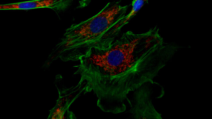
 and analyzed for confluency using AI (right).")
AI Confluency Analysis for Enhanced Precision in 2D Cell Culture
This article explains how efficient, precise confluency assessment of 2D cell culture can be done with artificial intelligence (AI). Assessing confluency, the percentage of surface area covered,…
Loading...

Precision and Efficiency with AI-Enhanced Cell Counting
This article describes the use of artificial intelligence (AI) for precise and efficient cell counting. Accurate cell counting is important for research with 2D cell cultures, e.g., cellular dynamics,…
Loading...
 of U2OS cells which were transfected with a fluorescently labelled protein. A fluorescence image of the cells (right) is also shown. The analysis and imaging were performed with Mateo FL.")
Leveraging AI for Efficient Analysis of Cell Transfection
This article explores the pivotal role of artificial intelligence (AI) in optimizing transfection efficiency measurements within the context of 2D cell culture studies. Precise and reliable…
Loading...
 where cyan indicates nuclei and magenta tight junctions.")
Rapid Check of Live Stem Cells in Cell-Culture Inserts set in Multi-Well Plates
See how efficient imaging of live iPSC stem cells within cell-culture inserts set in a multi-well plate can be done to evaluate the cells using a THUNDER Imager. Just read this article.
Loading...

Notable AI-based Solutions for Phenotypic Drug Screening
Learn about notable optical microscope solutions for phenotypic drug screening using 3D-cell culture, both planning and execution, from this free, on-demand webinar.
Loading...
Studying Virus Replication with Fluorescence Microscopy
The results from research on SARS-CoV-2 virus replication kinetics, adaption capabilities, and cytopathology in Vero E6 cells, done with the help of fluorescence microscopy, are described in this…
Loading...

How to Determine Cell Confluency with a Digital Microscope
This article shows how to measure cell confluency in an easy and consistent way with Mateo TL, increasing confidence in downstream experiments.
Loading...

How to do a Proper Cell Culture Quick Check
In order to successfully work with mammalian cell lines, they must be grown under controlled conditions and require their own specific growth medium. In addition, to guarantee consistency their growth…
Loading...

Deep Visual Proteomics Provides Precise Spatial Proteomic Information
Despite the availability of imaging methods and mass spectroscopy for spatial proteomics, a key challenge that remains is correlating images with single-cell resolution to protein-abundance…