
Science Lab
Science Lab
The knowledge portal of Leica Microsystems offers scientific research and teaching material on the subjects of microscopy. The content is designed to support beginners, experienced practitioners and scientists alike in their everyday work and experiments. Explore interactive tutorials and application notes, discover the basics of microscopy as well as high-end technologies – become part of the Science Lab community and share your expertise!
Filter articles
Tags
Products
Loading...

Understanding Clearly the Magnification of Microscopy
To help users better understand the magnification of microscopy and how to determine the useful range of magnification values for digital microscopes, this article provides helpful guidelines.
Loading...

ISO 9022 Standard Part 11 - Testing Microscopes with Severe Conditions
This article describes a test to determine the robustness of Leica microscopes to mold and fungus growth. The test follows the specifications of the ISO 9022 part 11 standard for optical instruments.
Loading...

Key Factors to Consider When Selecting a Stereo Microscope
This article explains key factors that help users determine which stereo microscope solution can best meet their needs, depending on the application.
Loading...

What is the FusionOptics Technology?
Leica stereo microscopes with FusionOptics provide optimal 3D perception. The brain merges two images, one with large depth of field and the other with high resolution, into one 3D image.
Loading...

Microscope Ergonomics
This article explains microscope ergonomics and how it helps users work in comfort, enabling consistency and efficiency. Learn how to set up the workplace to keep good posture when using a microscope.
Loading...
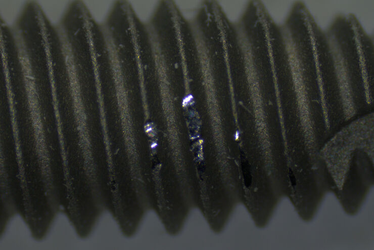
 taken with a ring light (RL) and near vertical illumination (NVI).")
Microscope Illumination for Industrial Applications
Inspection microscope users can obtain information from this article which helps them choose the optimal microscope illumination or lighting system for inspection of parts or components.
Loading...

Top Challenges for Visual Inspection
This article discusses the challenges encountered when performing visual inspection and rework using a microscope. Using the right type of microscope and optical setup is paramount in order to…
Loading...
Why is Manual Visual Inspection of Medical Devices so Challenging?
This article discusses how manual visual inspection, which is prevalent in the medical device industry, can lead to inconsistent results. It also addresses the challenges quality managers and…
Loading...

Image Gallery: THUNDER Imager
To help you answer important scientific questions, THUNDER Imagers eliminate the out-of-focus blur that clouds the view of thick samples when using camera-based fluorescence microscopes. They achieve…