
Science Lab
Science Lab
The knowledge portal of Leica Microsystems offers scientific research and teaching material on the subjects of microscopy. The content is designed to support beginners, experienced practitioners and scientists alike in their everyday work and experiments. Explore interactive tutorials and application notes, discover the basics of microscopy as well as high-end technologies – become part of the Science Lab community and share your expertise!
Filter articles
Tags
Products
Loading...
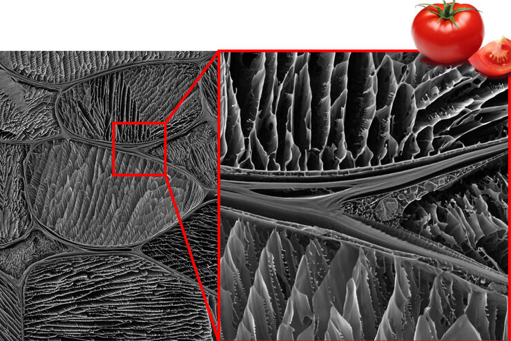
Studying the Microstructure of Natural Polymers in Fine Detail
The potential of cryogenic broad ion beam milling used in combination with scanning electron microscopy (cryo-BIB-SEM) for imaging and analyzing the microstructure of cryogenically stabilized soft…
Loading...

Expert Knowledge on High Pressure Freezing and Freeze Fracturing in the Cryo SEM Workflow
Get an insight in the working methods of the laboratory and learn about the advantages of Cryo SEM investigation in EM Sample Preparation. Find out how high pressure freezing, freeze fracturing and…
Loading...
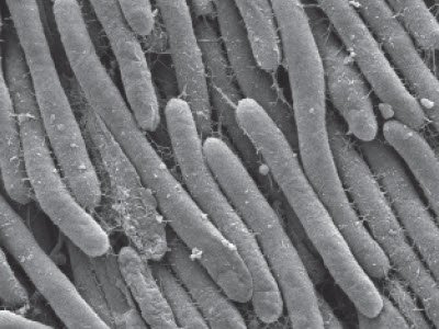
Bacteria Protocol - Critical Point Drying of E. coli for SEM
Application Note for Leica EM CPD300 - Critical point drying of E. coli with subsequent platinum / palladium coating and SEM analysis. Sample was inserted into a filter disc (Pore size: 16 - 40 μm)…
Loading...
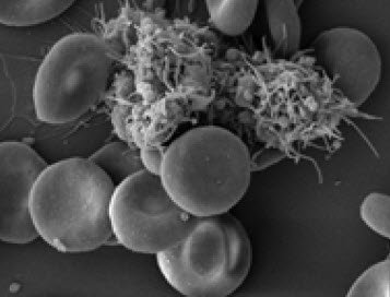
Human Blood Cells Protocol
Application Note for Leica EM CPD300 - Life Science Research. Species: Human (Homo sapiens)
Critical point drying of human blood with subsequent platinum / palladium coating and SEM analysis.
Loading...
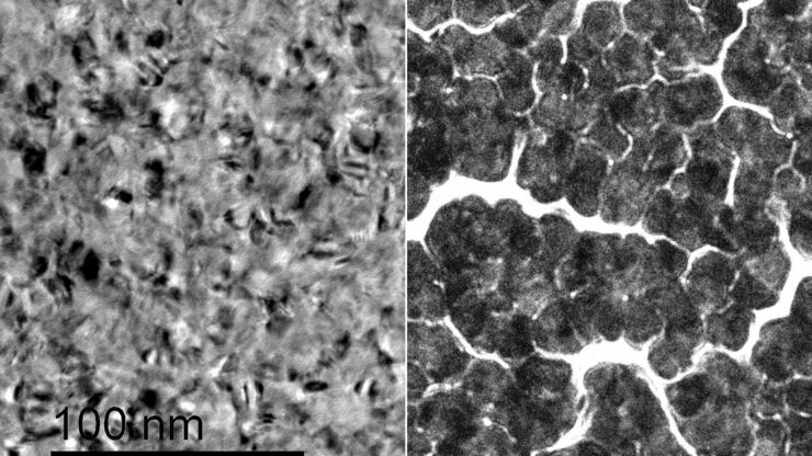
Improvement of Metallic Thin Films for HR-SEM by Using DC Magnetron Sputter Coater
Preparation techniques, like several kinds of coating methods play an important role for high resolution scanning electron microscopy (HR-SEM). Nonconductive sample like biological and synthetic…
Loading...
Brief Introduction to Freeze Fracture and Etching
Freeze fracture describes the technique of breaking a frozen specimen to reveal internal structures. Freeze etching is the sublimation of surface ice under vacuum to reveal details of the fractured…
Loading...
Brief Introduction to Coating Technology for Electron Microscopy
Coating of samples is required in the field of electron microscopy to enable or improve the imaging of samples. Creating a conductive layer of metal on the sample inhibits charging, reduces thermal…
Loading...
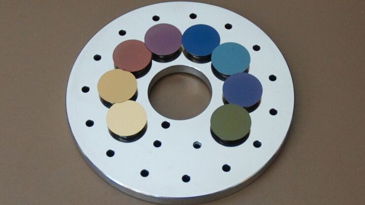
Carbon Thickness Evaluation in Electron Microscopy
The coating layers applied and used for electron microscopy imaging are commonly controlled and measured by quartz crystals. These crystals oscillate with a certain frequency (around 6 megahertz when…
