
Science Lab
Science Lab
The knowledge portal of Leica Microsystems offers scientific research and teaching material on the subjects of microscopy. The content is designed to support beginners, experienced practitioners and scientists alike in their everyday work and experiments. Explore interactive tutorials and application notes, discover the basics of microscopy as well as high-end technologies – become part of the Science Lab community and share your expertise!
Filter articles
Tags
Products
Loading...
, actin network (ATTO 647N), and nuclear pore basket (CF 680R).")
The Guide to STED Sample Preparation
This guide is intended to help users optimize sample preparation for stimulated emission depletion (STED) nanoscopy, specifically when using the STED microscope from Leica Microsystems. It gives an…
Loading...
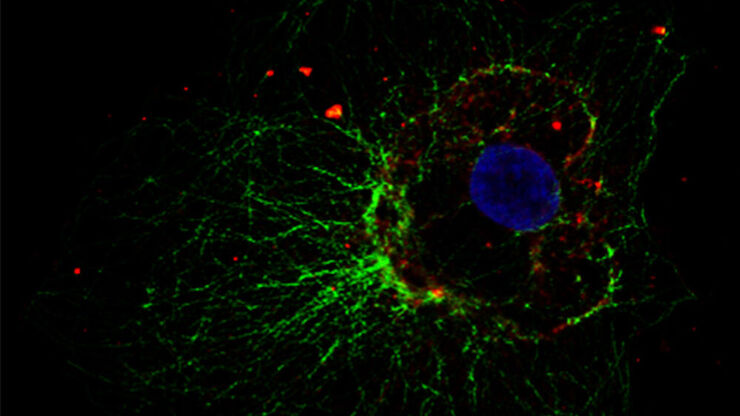
Studying Virus Replication with Fluorescence Microscopy
The results from research on SARS-CoV-2 virus replication kinetics, adaption capabilities, and cytopathology in Vero E6 cells, done with the help of fluorescence microscopy, are described in this…
Loading...

Methods to Improve Reproducibility in Spatial Biology Research
Establish reproducibility results for a Cell DIVE multiplexed imaging study in cancer research using the BAB 200 automated system from ASLS and validated antibodies from CST
Loading...
 tissue on a single slide.")
Characterizing tumor environment to reveal insights and spatial resolution
Antibodies from Cell Signaling Technology are validated for use with the Cell DIVE multiplexing workflow and used to probe cell lineages in the tumor microenvironment
Loading...

Dig Deeper Into the Complexities of Pancreatic Cancer with Multiplex Imaging
Cell DIVE is an iterative staining workflow for multiplexed imaging that unveils biological pathways to dig deeper into the complexities of pancreatic cancer.
Loading...

Deep Visual Proteomics Provides Precise Spatial Proteomic Information
Despite the availability of imaging methods and mass spectroscopy for spatial proteomics, a key challenge that remains is correlating images with single-cell resolution to protein-abundance…
Loading...

Multiplexed Imaging Types, Benefits and Applications
Multiplexed imaging is an emerging and exciting way to extract information from human tissue samples by visualizing many more biomarkers than traditional microscopy. By observing many biomarkers…
Loading...
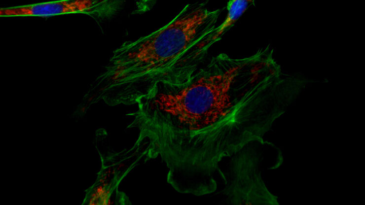
How to Prepare your Specimen for Immunofluorescence Microscopy
Immunofluorescence (IF) is a powerful method for visualizing intracellular processes, conditions and structures. IF preparations can be analyzed by various microscopy techniques (e.g. CLSM,…
Loading...
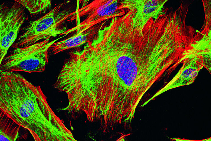
Fluorescent Dyes
A basic principle in fluorescence microscopy is the highly specific visualization of cellular components with the help of a fluorescent agent. This can be a fluorescent protein – for example GFP –…