
Science Lab
Science Lab
The knowledge portal of Leica Microsystems offers scientific research and teaching material on the subjects of microscopy. The content is designed to support beginners, experienced practitioners and scientists alike in their everyday work and experiments. Explore interactive tutorials and application notes, discover the basics of microscopy as well as high-end technologies – become part of the Science Lab community and share your expertise!
Filter articles
Tags
Products
Loading...
 and analyzed for confluency using AI (right).")
AI Confluency Analysis for Enhanced Precision in 2D Cell Culture
This article explains how efficient, precise confluency assessment of 2D cell culture can be done with artificial intelligence (AI). Assessing confluency, the percentage of surface area covered,…
Loading...

Precision and Efficiency with AI-Enhanced Cell Counting
This article describes the use of artificial intelligence (AI) for precise and efficient cell counting. Accurate cell counting is important for research with 2D cell cultures, e.g., cellular dynamics,…
Loading...
 of U2OS cells which were transfected with a fluorescently labelled protein. A fluorescence image of the cells (right) is also shown. The analysis and imaging were performed with Mateo FL.")
Leveraging AI for Efficient Analysis of Cell Transfection
This article explores the pivotal role of artificial intelligence (AI) in optimizing transfection efficiency measurements within the context of 2D cell culture studies. Precise and reliable…
Loading...

Overcoming Observational Challenges in Organoid 3D Cell Culture
Learn how to overcome challenges in observing organoid growth. Read this article and discover new solutions for real-time monitoring which do not disturb the 3D structure of the organoids over time.
Loading...

How to Streamline Your Histology Workflows
Streamline your histology workflows. The unique Fluosync detection method embedded into Mica enables high-res RGB color imaging in one shot.
Loading...
Accelerating Discovery for Multiplexed Imaging of Diverse Tissues
Explore IBEX: Open-source multiplexed imaging. Join the collaborative IBEX Imaging Community for optimized tissue processing, antibody selection, and human atlas construction.
Loading...

Notable AI-based Solutions for Phenotypic Drug Screening
Learn about notable optical microscope solutions for phenotypic drug screening using 3D-cell culture, both planning and execution, from this free, on-demand webinar.
Loading...

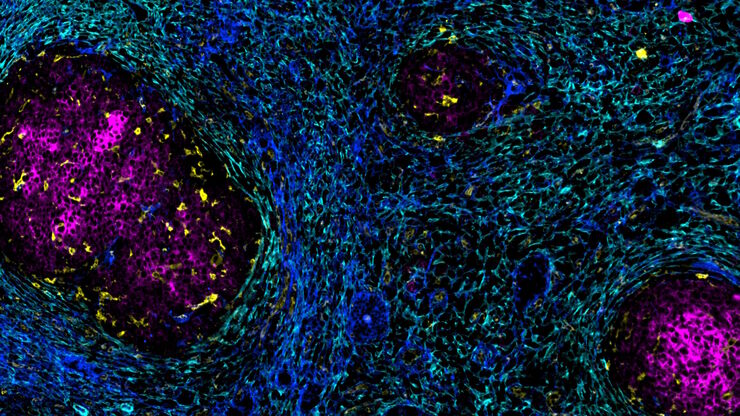
Understanding Tumor Heterogeneity with Protein Marker Imaging
Explore tumor heterogeneity and immune cell dynamics. See how quantitative imaging analysis reveals spatial relationships and molecular insights crucial for advancing cancer research and therapeutics.
Loading...

In Situ Identification of Cancer Stem Cell Niches in Hepatocellular Carcinoma
Discover how multiplexed imaging technology uncovers cancer stem cell niches in Hepatocellular Carcinoma using multiplex immunodetection, revealing extracellular matrix dynamics. Explore precise…