Cell Culture
Get what you need and increase the efficiency of your imaging workflow for cell and tissue culture with inverted microscopes from Leica Microsystems.
These easy-to-use microscopes allow you to configure an imaging solution which is optimal for your needs. They have flexible options for the condenser lens and digital imaging documentation features, creating a solution that is just right for your lab.
Our experts on solutions for cell-and-tissue-culture applications are happy to help you with their advice.
Leica cell & tissue culture microscopes feature
Easy-to-use operation
Easy-to-use operation that requires minimal training and maintenance so you can fully concentrate on your research
Cool, color-safe LED illumination
Cool, color-safe LED illumination for constant color temperature through all stages of intensity
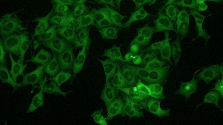
Easy fluorescence
Easy fluorescence (optional) to easily visualize your fluorescent markers
HD imaging
HD imaging (optional) – connect the HD camera directly to a monitor or PC; providing high quality publication images
Flexible working distance
Flexible working distance up to 80 mm for accommodating slides, petri dishes, multi-well dishes, and taller flasks
Cell factory
Cell factory solution fits vessels up to 400 mm tall
Microscopes – Basic Requirements
What tools do I need for cell and tissue culture?
To manage the daily work of a cell-and-tissue-culture lab, a microscope with an inverse configuration is needed. Such an inverted microscope features the objective lens below and the condenser above the specimen, enabling the objective to be placed in proximity of the cells, often at the bottom of flasks, and a large working distance above the specimen so it can be easily handled during imaging.
Due to the very low intrinsic contrast of animal cells, a cell-and-tissue-culture microscope has to deliver contrast methods like phase contrast. DIC (Differential Interference Contrast) doesn’t help here, because it does not work with the plastic vessels used for cell and tissue culture. A very good alternative for DIC is IMC (Integrated Modulation Contrast), which works with plastic containers and, in addition, doesn’t need special objectives or prisms. Moreover, a cell-and-tissue-culture microscope should be easy to handle so cell imaging can be done efficiently.
Leica cell-and-tissue-culture microscopes offer you the ease of use and flexibility with contrast methods that you need for your individual requirements.
Related Articles

History, Developments and Trends of Microscopy in Cancer Research

Researchers Insights: Microscopy in Cancer Research

AI meets Deep Visual Proteomics (DVP) to Advance Disease Research
 and co-stained for nuclear DNA (Hoechst 33342), microtubules (Alexa 555) and F-actin (ATTO 643). Image was captured on Mateo FL.")
Microscopy and AI Solutions for 2D Cell Culture
.")
AI-Powered Hi-Plex Spatial Analysis Tools for Breast Cancer Research
.")
Focus on Long-Term Imaging in 3D with Light Sheet Microscopy

Volume EM and AI Image Analysis
 at Leica Microsystems.")
How a Breakthrough in Spatial Proteomics Saved Lives
 at 2 weeks. Image acquired using Mica.")
How to Image Axon Regeneration in Deep Muscle Tissue

Capturing Developmental Dynamics in 3D
Tutorials Basic Microscopy
Phase Contrast
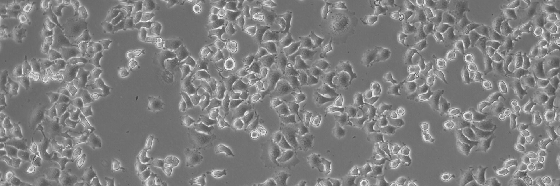
Phase contrast is an optical contrast technique for making unstained phase objects (e.g. flat cells) visible under the optical microscope. Cells that appear inconspicuous and transparent in brightfield can be viewed in high contrast and rich detail using a phase contrast microscope.
Differential Interference Contrast
Differential interference contrast (DIC) microscopy is a good alternative to brightfield microscopy for gaining proper images of unstained specimens that often only provide a weak image in brightfield.
Integrated Modulation Contrast
Hoffman modulation contrast has established itself as a standard for the observation of unstained, low-contrast biological specimens. Its innovative technical implementation permits significantly simpler handling and greater flexibility in deployment.
Find your Cell Culture Solution
When it comes to cell and tissue cultures, there are some important differences between the solutions. To help you find the appropriate solution for your cell-and-tissue-culture needs, please answer these three quick questions.
Contact us{{ question.questionText }}
Please select an answer!
Best Match
{{ resultProduct.header }}
{{ resultProduct.subheader }}
{{ resultProduct.description }}
{{ resultProduct.features }}
Request Your Information Package
Our Basic Cell Culture Products
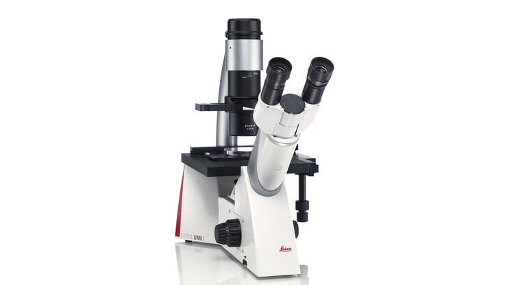
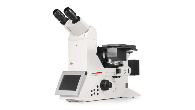
Inverted Microscope for Cell Culture Leica DMi1
The Leica DMi1 inverted microscope supports your specific work routine in your cell culture lab. Its operation is so intuitive, its handling so comfortable that you can fully concentrate on your work. Choose the functions you need, and, if necessary, you can easily add a variety of accessories that are important for your work.
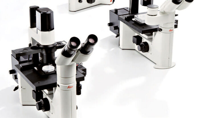
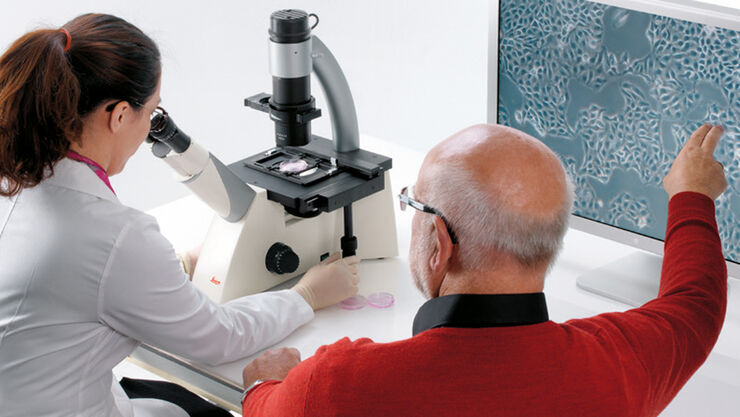
Inverted Laboratory Microscope Leica DM IL LED
The Leica DM IL LED features a comprehensive set of contrast methods to monitor your specimen the way you need. High-quality Phase contrast, excellent modulation contrast and brilliant fluorescence are just one fingertip away. Robust stability, plenty of space to work with tools, long working distances to accommodate large culture flasks and a stable illumination without heat make work at the microscope easy and convenient.
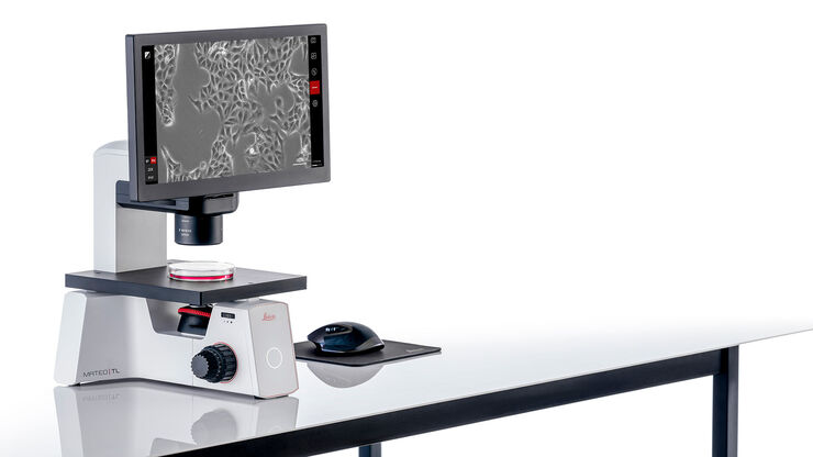
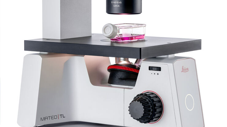
Digital Transmitted Light Microscope Mateo TL
For researchers who need consistent experimental outcomes, Mateo TL enables all lab members to check and document cell growth status conveniently and comfortably. Measure confluency consistently, thereby increasing the confidence in the success of their downstream experiments.
Brightfield | Phase contrast | DIC | IMC | Fluorescence | Magnification | Working Distance | Camera | |
Leica DM IL LED | + | + | - | + | + | PH: 5x to 63x IMC: 10x, 20x, 32x, 40x | 40 mm, 80 mm | + (free choice) |
Leica DMi1 | + | + | - | - | - | 10x, 20x, 40x | 40 mm, 50 mm, 80 mm | + (integrated) |
Mateo TL | + | + | - | - | - | 4x, 10x, 20x, 40x | 50 mm | + (integrated) |
Mateo FL | + | + | - | - | + | 2.5x, 4x, 5x, 10x, 20x, 40x, 63x | 50 mm | + dual camera (integrated) |
Microscopes dedicated to cell-and-tissue-culture lab work.
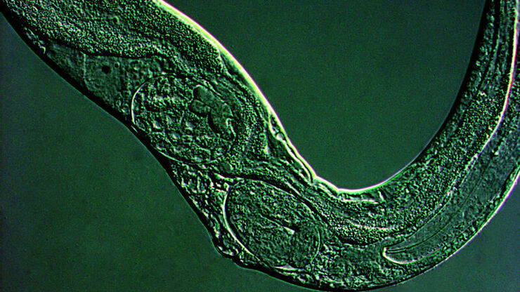
How to Grow Cells
Animal cells are cultured in all kinds of different vessels, ranging from tiny microfluidic devices used for basic research to 96-well plates for screening, cell-and-tissue-culture flasks, and cell factories for large scale pharmaceutical production.
Due to their disposable use, the majority of containers is made of plastic. Others are specifically adopted to microscopy applications and therefore have a glass bottom.
The medium for animal cell and tissue culture contains:
- water;
- an energy source;
- amino acids;
- vitamins; and
- salts.
In addition to water and nutrients, it also includes a buffer system plus a pH indicator which can check if the cell medium is maintaining a balanced pH level.
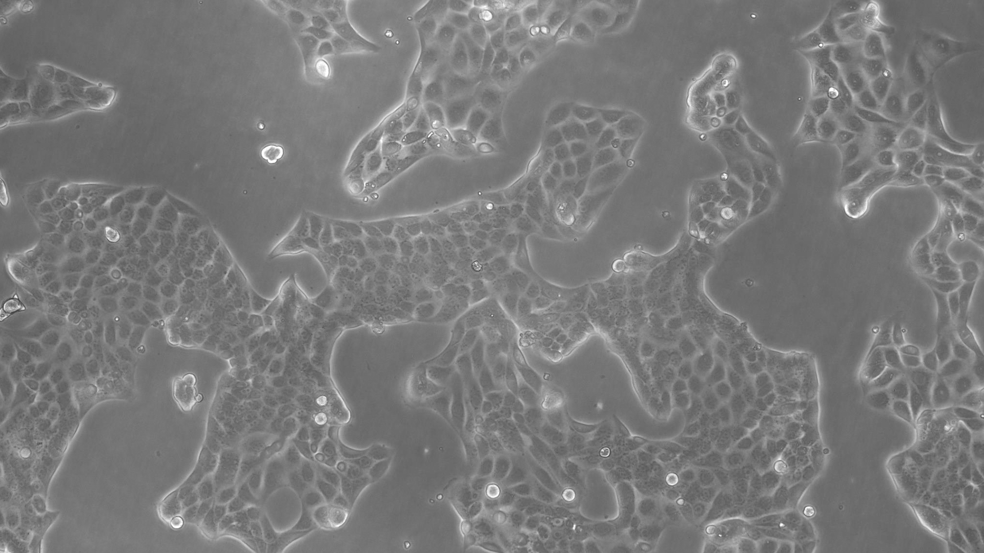
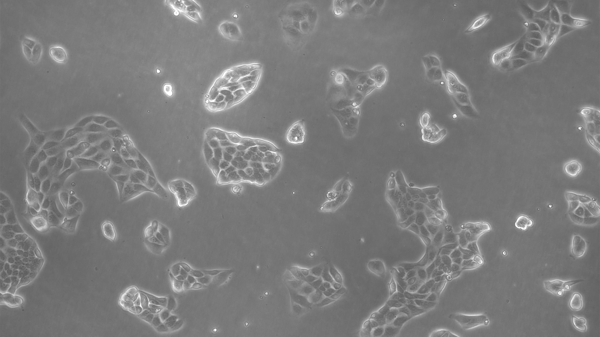
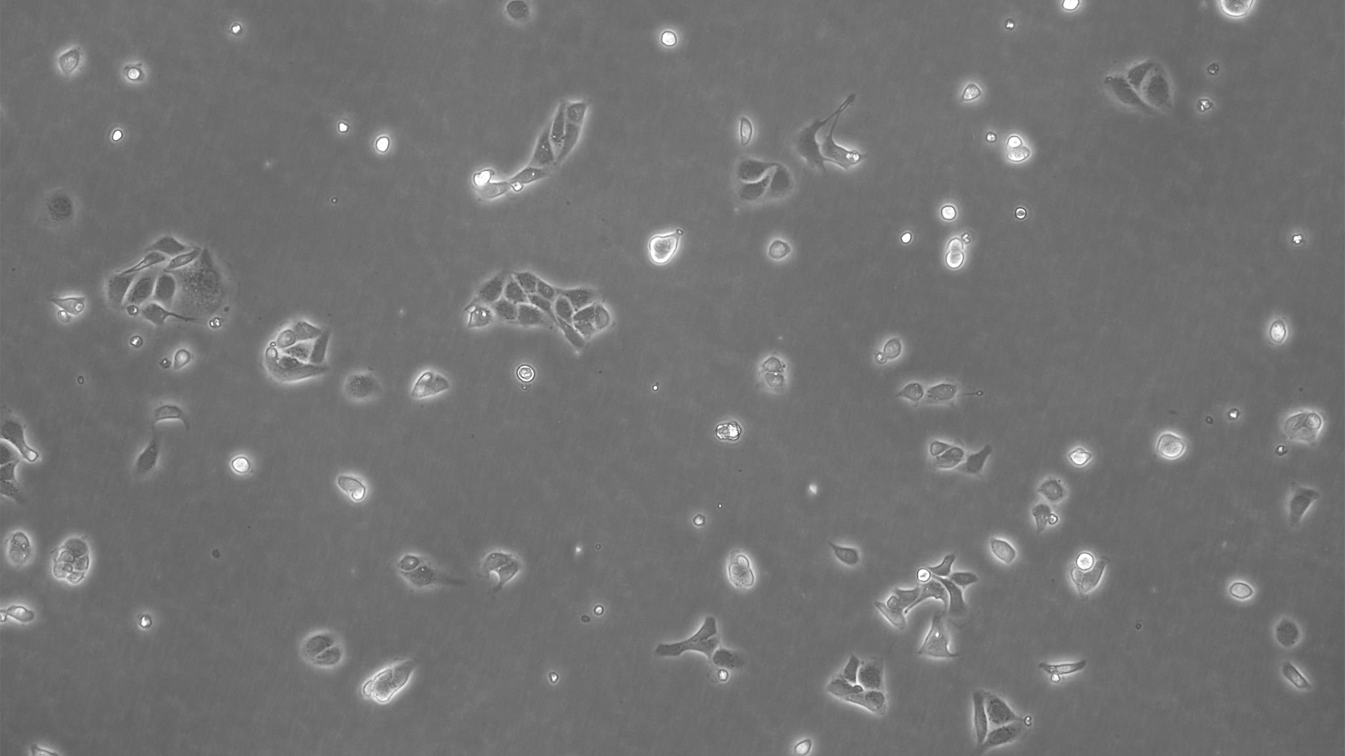
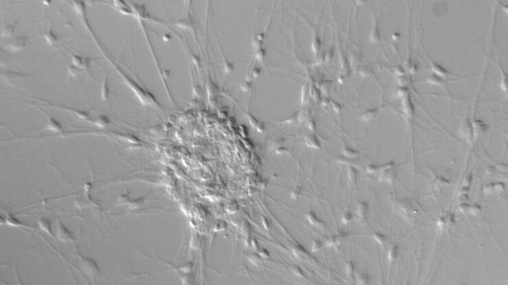
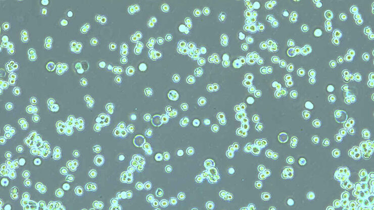
The Appearances of Cells
Lab-grown animal cells can be distinguished due to several criteria:
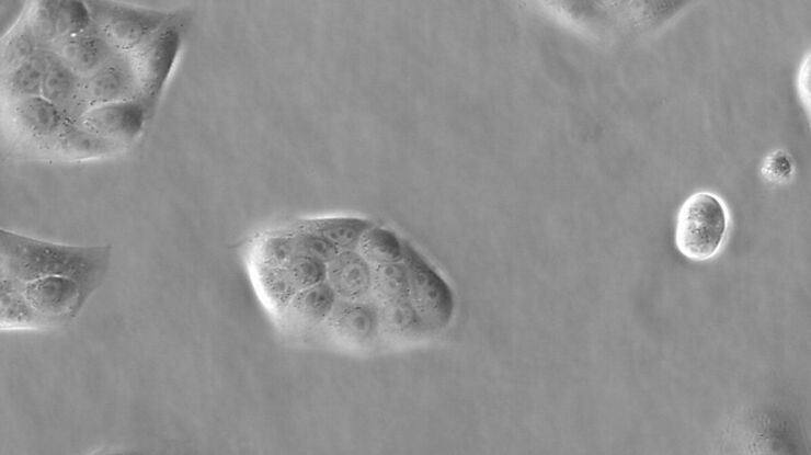
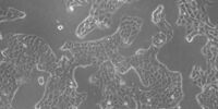
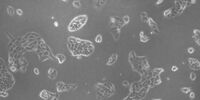
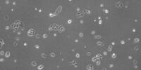
Their morphology is easy to identify with the microscope. Whereas fibroblast-like cells have a bi- or multi-polar, elongated shape, epithelial-like cells show a polygonal outline. In contrast to these first 2 cell types, lymphoblast-like cells don’t grow by adhering to a surface, but in suspension;
The type of cell can be subdivided into immortalized cells, primary cells, and stem cells;
The cell organization can range from simple 2D mono-culture to 2D co-culture as well as 3D spheroids and organoids.
.")
Name | Morphology | Source |
COS | Fibroblast-like | African green monkey |
HEK 293 | Epithelial-like | Human |
CHO | Epithelial-like | Hamster |
MDCK | Epithelial-like | Dog |
HeLa | Fibroblast-like | Human |
Jurkat | Lymphoblast-like | Human |
A few examples of cell lines used for cell and tissue culture.
Microscopes – Advanced Requirements
What tools do I need?
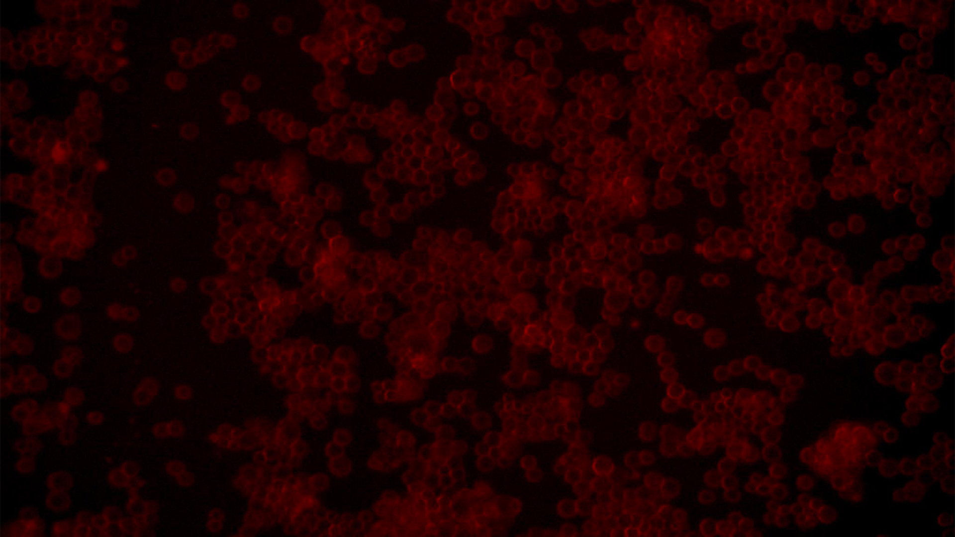
A very common approach is to transfect cells with fluorescent markers for subsequent investigation with a research optical microscope. If you are dealing with fluorescent proteins, also your cell-and-tissue-culture microscope needs a fluorescence option which allows transfection efficiency to be checked.
For meaningful documentation and standardization of work, the microscope should have a digital camera for the recording of acquired image data which can be further analyzed.
As space is an issue in many labs, a cell-and-tissue-culture microscope shouldn’t be too big, otherwise it may not be able to fit in a fume or flow hood. Moreover, recent trends demand microscopes which are small and robust enough to be used even inside an incubator.
Inverted Microscope Solution DMi8 S Platform
Whether you need to precisely follow the development of a single cell in a dish, screen through multiple assays, obtain single molecule resolution, or tease out behaviors of complex processes, a DMi8 S system will enable you to see more, see faster, and find the hidden.
Frequently Asked Questions Cell Culture
For you to have an overview on the overall status of a cell and tissue culture, a magnification of 100x – 200x is sufficient. The low intrinsic contrast of mammalian cells and tissues means specific contrast methods are needed to clearly observe them. Simple brightfield microscopy is often not sufficient. Phase contrast and integrated modulation contrast (IMC) are the most common ones which enable clear visualization of cells and tissues, whereas differential interference contrast (DIC) is not compatible with the plastic vessels normally used for cell and tissue culture. For fluorescence microscopy, the cells or tissues have to be transfected or labelled with fluorescent markers prior to imaging. For more information, refer to the article: Introduction to Mammalian Cell Culture
Cells are able to adhere to other cells and biocompatible surfaces due to interactions between cell-adhesion molecules, proteins in the cell plasma membrane, and other proteins or polypeptides. The bottom surface of plastic flasks and plates used for cell and tissue culture are treated and then coated with any number of a variety of biomaterials, such as collagen, laminin, fibronectin, heparin sulfate, hyaluronidate, etc. Synthetic polypeptides, like poly-lysine, have also been used as coatings for the plastic surfaces, because it creates a positive charge which can enhance cell attachment.
Useful Tutorials
How to do a Proper Cell Culture Quick Check
Many fields of biomedical research, like cancer research, drug development and tissue engineering, require the use of living cells to perform a variety of assays. Mammalian cell cultures are an essential tool in biology because they allow rapid growth and proliferation of different cell types for experimental analysis.
Fluorescent Proteins
The prospects of fluorescence microscopy changed dramatically with the discovery of fluorescent proteins in the 1950s. The starting point was the detection of the jellyfish Aequorea victoria green fluorescent protein (GFP) by Osamo Shimomura. Hundreds of GFP mutants later, the range of fluorescent proteins reaches from the blue to the red spectrum.
An Introduction to Fluorescence
Fluorescence is an effect which was first described by George Gabriel Stokes in 1852. He observed that fluorite begins to glow after being illuminated with ultraviolet light. Fluorescence is a form of photoluminescence which describes the emission of photons by a material after being illuminated with light. The emitted light is of longer wavelength than the exciting light. This effect is called the Stokes shift.
