
Clinical Pathology Microscope Solutions
In clinical microscopy, accurate and timely diagnoses are critical in the management and treatment of medical patients. Efficiency therefore plays an important role in the daily routine of clinical pathologists.
Microscopes can help pathologists achieve efficiency in four ways:
- Enabling an easy customizable setup up and operation
- Making it easy to provide high-quality images that facilitate accurate diagnoses.
- Working with cameras that offer TWAIN capability to archive images in a hospital or lab information system.
- Creating ergonomic laboratory workstations that reduce strain and tension for clinicians that spend many hours on the microscope each day.
Contact us to learn how Leica clinical microscopes can support timely, accurate diagnoses.


What is clinical pathology?
Clinical pathology is concerned with the diagnosis of infections and disease based on the laboratory analysis, measurement, and quantification of bodily fluids, tissues, and microorganisms. Laboratory medicine is a term often used synonymously with clinical pathology.

What are microscopes used for in clinical pathology?
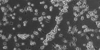
Clinical pathologists use microscopes to look at bodily fluids like blood, lymph, etc. to find abnormalities. Medical microbiologists, also called clinical microbiologists, look at microorganisms, such as bacteria, fungi, and parasites. Microscopes magnify details in the specimens and microorganisms and, thus, make visible information which could not be discerned with the naked eye.

What kind of microscopes do clinical pathologists use?
Most clinical pathology labs use bright-field microscopes to examine their samples, as this type of microscope usually provides high enough resolution with an illuminated background that helps them to see their area of interest more easily. However, some applications sometimes require more specialized microscope solutions that offer higher performance, such as fluorescence and laser dissection microscopy.
Our Solutions for Clinical Pathology
DM1000 LED | Visoria B | DM3000 & DM3000 LED | |
| Occasional use | x | - | - |
| Average to high workload | - | x | x |
| Adjustable both microscope height and viewing height & angle with eyepieces | x | x | x |
| Control knobs in close proximity with symmetrical layout | - | x | x |
| Adjustable height of stage and focus controls | x | x | x |
| Adjustable torque for focus knobs | - | x | x |
| Multiple viewing system for simultaneous observation by several users | - | x | - |
| Fast objective change with toggle mode | - | - | x |
| Automated illumination manager | - | x | x |
| Robust compact design | x | x | x |
| Go to product page | Go to product page | Go to product page |
x = included, - = not available
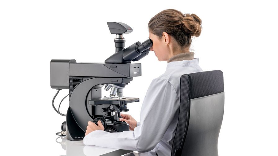
Why do ergonomics matter in clinical pathology?
Many pathologists work long hours with their microscopes, and this means their necks and backs may be physically strained from standing or sitting at an unnatural angle. Hand and arm muscles may feel tense due to small, repetitive movements for focusing and moving the stage. Eyes may also become strained by the microscope illumination.
Musculoskeletal pain may have serious effects. It can impact pathologists’ well-being and quality of life, as well as their productivity and diagnostic efficiency.
Leica clinical microscopes can support accurate and timely diagnoses as they make workflows more efficient with their ergonomic design and excellent optics.
I am working with Leica microscopes since my pathology training at Ghent University. When using or testing other microscopes, they never have the image quality and ergonomic features that Leica microscopes provide.
The role of microscopes in hematology
Hematology is the subset of clinical pathology that concentrates on studying blood-related disorders. It encompasses the examination of blood cells, coagulation factors, and bone marrow.
Microscopy plays a pivotal role by enabling the visualization of blood smears and bone marrow aspirates, aiding in the identification of abnormalities, such as anemia, leukemia, and clotting disorders. This microscopic analysis provides important insights into hematologic conditions, guiding diagnosis, treatment decisions, and monitoring of patient responses to therapy.


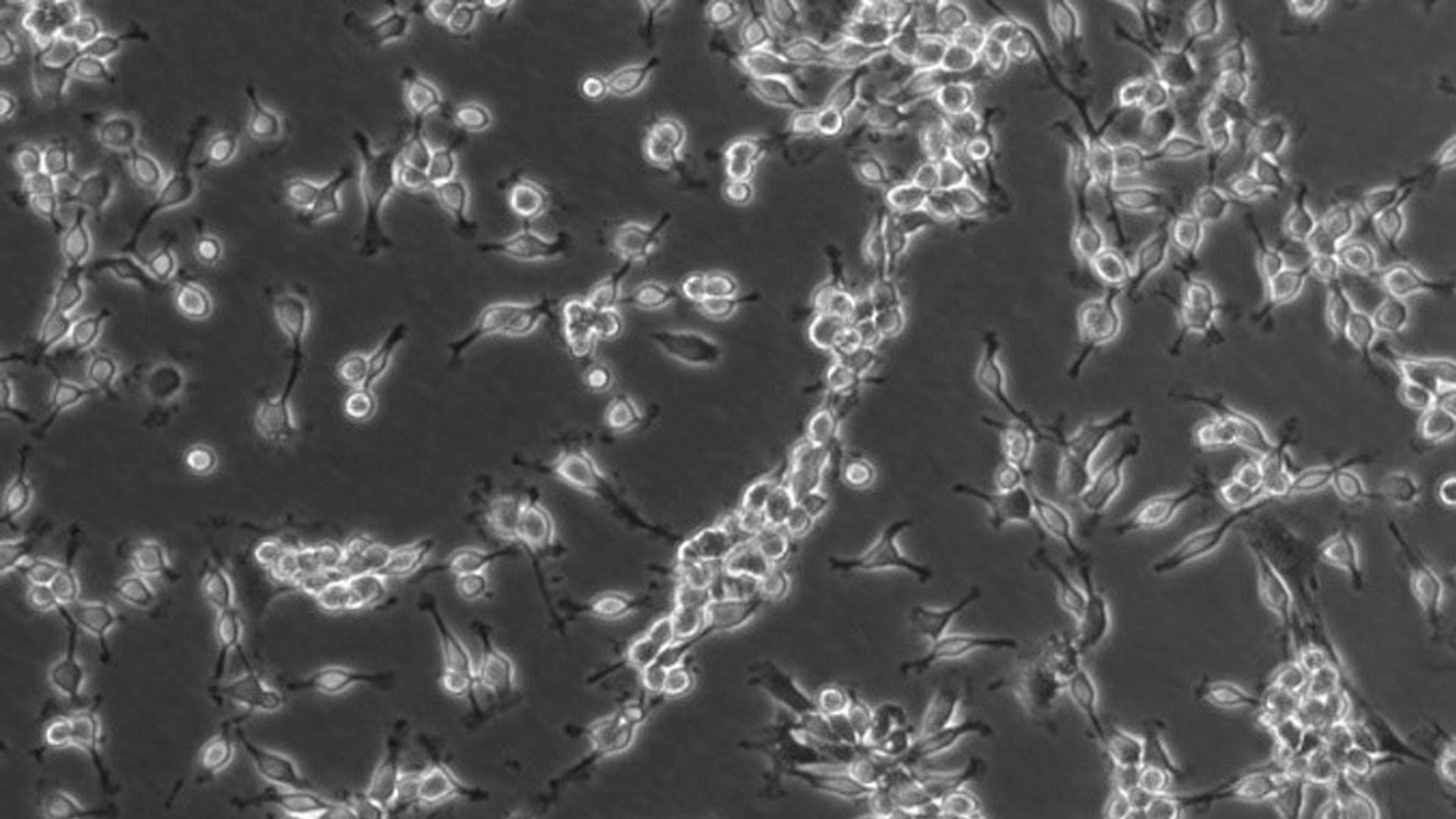
Discovering the hidden in clinical microbiology
Clinical microbiology is a discipline involving the study of microscopic organisms like bacteria, viruses, fungi, and parasites within biological samples to help diagnose and combat infectious diseases. Various types of microscopies, including light electron (EM) and fluorescence microscopes, can be used to visualize these microorganisms.
Advanced Leica microscope solutions enable microbiologists to continually take their work further by identifying pathogens, studying their structures, behaviors, and interactions with host cells, facilitating new discoveries. This microscopic analysis helps improve patient outcomes by enabling timely interventions in medical treatment and the development of targeted therapies against infectious diseases.
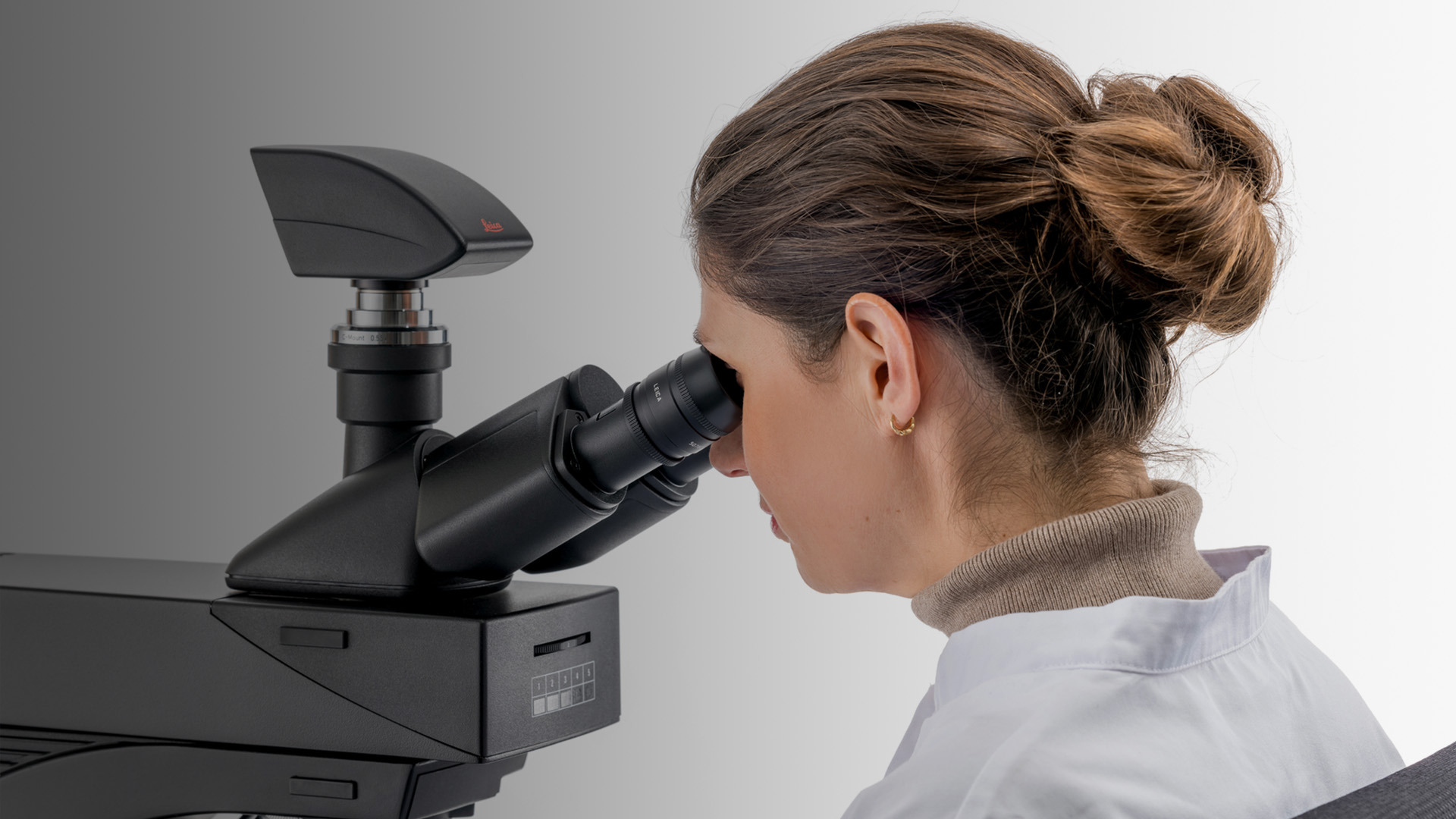
How comfortable is your microscope?
Your clinical microscope should be adaptable to your needs, not vice versa. To keep physical strain to a minimum, you should be able to:
- Adjust the height of the microscope to your own height.
- Choose the right eyepieces and viewing angle to achieve a comfortable position.
- Rest your arms and hands on the workbench, with the focus knobs within comfortable reach.
- Easily keep a symmetrical position with aligned shoulders without leaning to the side.
- Avoid repetitive movements through automated features.
- Balance the light intensity, even when you change objectives, to help reduce the risk of eye strain.
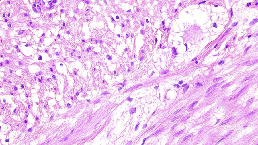
How does image quality support accurate diagnoses?
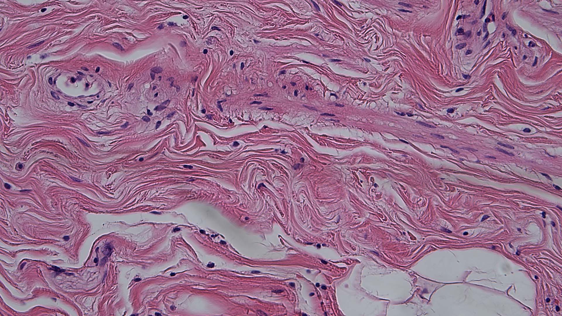
Pathologists need to discern subtle color differences, particularly in stained histological specimens such as Hematoxylin and Eosin (H&E) or papanicolaou for pap smears, to be able to make accurate and reliable diagnoses. After a quick overview, changing to a higher magnification should clearly show the more details.
For the particularly high magnifications used in clinical microbiology examinations, oil-immersion objectives are needed to discern details in the specimen. Only high-quality optics enable pathologists to see a clear, crisp image of their specimen so that they can draw the right conclusions from what they observe.
Additionally, with microscope cameras which have TWAIN capability, you can easily store and transfer your images directly to the hospital or laboratory information system, facilitating the creation of reports.
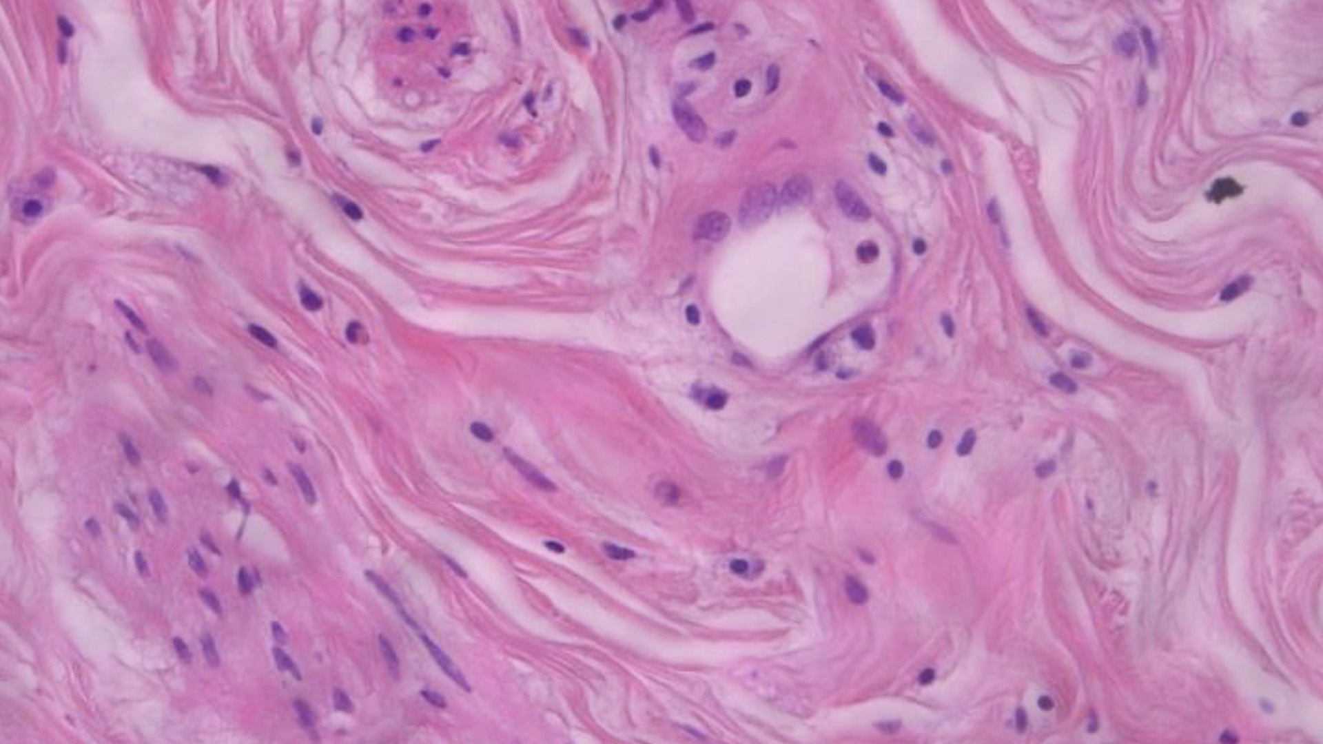
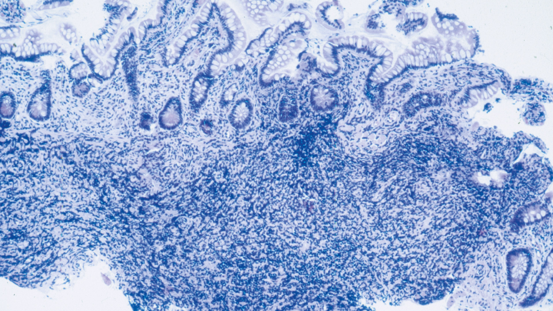
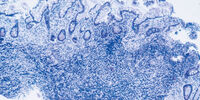
Differentiating shades of blue and purple
In clinical pathology, technicians often find themselves looking at samples with many similar color shades, making it difficult to see the details. Leica color-corrected optics will give you an image with clearly discernible areas which are free from color fringes, unlike objectives without correction. With the high color fidelity of color-corrected objectives you can be sure that the colors you see through the eyepieces are the exact colors that are present in the specimen.
Leica optics are world-renowned for their image quality and will serve you well, whether you use conventional or oil-immersion objectives. Cameras suitable for Leica pathology microscopes will also reproduce colors present in your specimen accurately. Their large bit depth and high dynamic range are designed to truthfully represent the different colors which are present in your specimen.
Frequently Asked Questions Clinical Pathology
The automated toggle mode of the DM3000 microscope allows users to switch from one objective to another using two buttons which are located near the focus knobs. Any two objectives can be chosen, so that users can switch quickly between low and high magnification, thus, seeing either an overview of the sample or a specific area of interest in detail.
With ergonomic microscopes, pathologists can work more comfortably and with less interruptions. An upright posture and a symmetrical setup of the microscope helps avoid strain in the back, neck, and shoulders, while ergonomic armrests and adjustable focus knobs prevent muscle tension in the hands and arms. Thus, pathologists can fully concentrate on their work and need fewer breaks to relax their muscles.
Yes, we offer DM Multiple Viewing Systems for up to 12 viewing stations.


